Назад

A comprehensive guide to noninvasive ventilation (NIV)

Статья

Автор: Karjaghli Munir, Respiratory Therapist, Hamilton Medical Clinical Application Specialist

Дата: 01.03.2024

Learn everything you need to know in our guide to NIV ventilation: from the medical abbreviation, to what noninvasive ventilation is, when to use it, selecting the interface and settings, and more.

A comprehensive guide to noninvasive ventilation (NIV)

Terminology, symbols, and abbreviations

Noninvasive ventilation is commonly referred to as NIV.

The term noninvasive positive-pressure ventilation (abbreviated NPPV or NIPPV) was previously used to distinguish it from noninvasive negative-pressure ventilation, but given the latter's rarity nowadays, the simpler term NIV is more convenient. As there is now a range of ventilators available for NIV, use of the product name BIPAP (Bilevel positive airway pressure) as a generic term for NIV should be avoided.

Other abbreviations are explained directly in the text.

Introduction

Noninvasive positive-pressure ventilation involves the delivery of oxygen into the lungs via positive pressure without the need for endotracheal intubation. It is used in both acute and chronic respiratory failure, but requires careful monitoring and titration to ensure its success and avoid complications (Rochwerg B, Brochard L, Elliott MW, et al. Official ERS/ATS clinical practice guidelines: noninvasive ventilation for acute respiratory failure. Eur Respir J. 2017;50(2):1602426. Published 2017 Aug 31. doi:10.1183/13993003.02426-20161​).

Over the past century, noninvasive ventilation (NIV) has improved dramatically and been used to treat respiratory failure from multiple etiologies. It has been proven more effective in preventing intubation compared to standard oxygen therapy in the acute setting (Gong Y, Sankari A. Noninvasive Ventilation. In: StatPearls. Treasure Island (FL): StatPearls Publishing; December 11, 2022.2​).

Respiratory support may be delivered using continuous positive airway pressure (CPAP) devices or those that deliver bilevel positive airway pressure (pressure-support ventilation, PSV). For the purposes of this article, the name NIV covers both CPAP and PSV (Gong Y, Sankari A. Noninvasive Ventilation. In: StatPearls. Treasure Island (FL): StatPearls Publishing; December 11, 2022.2​).

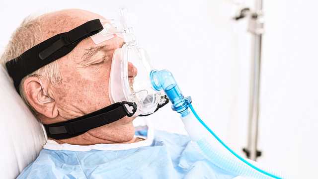
Adult with pulmodyne NIV mask
Illustration for the chapter "What are the benefits of NIV?"

What are the benefits? Goals and benefits of using NIV

We can divide the goals and benefits of NIV into the acute and long-term care setting as follows.

Benefits of NIV in acute care settings (Egan's fundamentals of respiratory care e-book3​):

  • Improve gas exchange
  • Avoid intubation
  • Decrease mortality
  • Decrease length of time on the ventilator
  • Decrease duration of hospitalization
  • Decrease incidence of ventilator-associated pneumonia
  • Relieve symptoms of respiratory distress
  • Improve patient-ventilator synchrony
  • Maximize patient comfort


Benefits of NIV in long-term care setting (Egan's fundamentals of respiratory care e-book3​):

  • Relieve or improve symptoms
  • Enhance quality of life
  • Avoid hospitalization
  • Increase survival
  • Improve mobility
Illustration for the chapter "How does NIV work?"

How does NIV work?

Noninvasive ventilation works by creating positive airway pressure, i.e., the pressure outside the lungs is greater than the pressure inside the lungs. This causes air to be forced into the lungs (down the pressure gradient), lessening the respiratory effort and reducing the work of breathing.

It also helps to keep the chest and lungs expanded by increasing the functional residual capacity (the amount of air remaining in the lungs after expiration) after normal (tidal) expiration; this is the air in the alveoli available for gas exchange (Rochwerg B, Einav S, Chaudhuri D, et al. The role for high flow nasal cannula as a respiratory support strategy in adults: a clinical practice guideline. Intensive Care Med. 2020;46(12):2226-2237. doi:10.1007/s00134-020-06312-y4​).

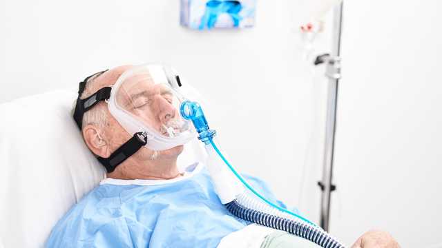
Adult with pulmodyne NIV mask

NIV modes

The following modes are noninvasive: 

  • CPAP: Stands for continuous positive airway pressure
  • NIV (PSV): Also known as bilevel CPAP mode, or bilevel pressure mode
  • NIV-ST (spontaneous/timed, S/T): Stands for spontaneous/timed noninvasive ventilation

CPAP

CPAP aims to deliver one continuous level of positive pressure throughout both the inspiratory and expiratory phases of breathing.

It improves oxygenation by opening collapsed airways, improving functional residual capacity (FRC), and improving preload and afterload in cardiogenic pulmonary edema (Bello G, De Santis P, Antonelli M. Non-invasive ventilation in cardiogenic pulmonary edema. Ann Transl Med. 2018;6(18):355. doi:10.21037/atm.2018.04.395​).

CPAP improves lung compliance and therefore reduces the effort required for breathing by preventing alveolar collapse and counteracting the excessive intrinsic PEEP seen in obstructive lung conditions such as COPD (Bello G, De Santis P, Antonelli M. Non-invasive ventilation in cardiogenic pulmonary edema. Ann Transl Med. 2018;6(18):355. doi:10.21037/atm.2018.04.395​, Davidson AC, Banham S, Elliott M, et al. BTS/ICS guideline for the ventilatory management of acute hypercapnic respiratory failure in adults [published correction appears in Thorax. 2017 Jun;72 (6):588]. Thorax. 2016;71 Suppl 2:ii1-ii35. doi:10.1136/thoraxjnl-2015-2082096​).

Good to know: When ΔPsupport /∆Pinsp is set to zero in NIV and NIV-ST, the ventilator functions like a conventional CPAP system.
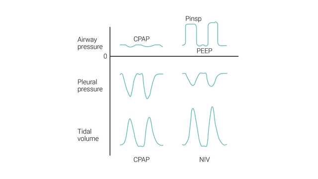
Illustration of CPAP and NIV
Adapted from Hess, Dean R., and Robert M. Kacmarek. Essentials of mechanical ventilation. McGraw Hill Education, 2019.
Illustration of CPAP and NIV
Adapted from Hess, Dean R., and Robert M. Kacmarek. Essentials of mechanical ventilation. McGraw Hill Education, 2019.

NIV (PSV)

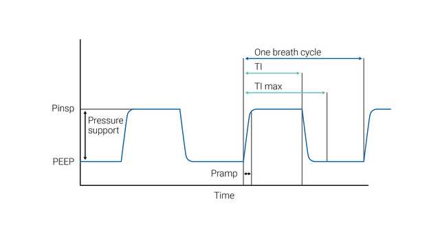
NIV (PSV) aims to deliver two levels of positive airway pressure support. The lower level is similar to CPAP; however, it is more commonly called positive end-expiratory airway pressure (PEEP) as it is present only at the expiratory phase of breathing. 

The patient’s inspiratory effort is assisted by the ventilator at a preset level of inspiratory pressure (ΔPsupport). Inspiration is triggered and cycled by the patient's effort. During NIV, the patient determines the respiratory rate, inspiratory time, and tidal volume.

The size of the breath (tidal volume) generated in a particular patient is dependent on the ΔPsupport setting – the higher the ΔPsupport setting, the greater the tidal volume.

ETS (expiratory trigger sensitivity) determines the spontaneous inspiratory time by cycling to expiration once the inspiratory flow decreases to a preadjusted percentage of the peak inspiratory flow.

In case the ETS criteria are not met (leakage), the inspiratory time can also be limited by TI max (maximum inspiratory time). 
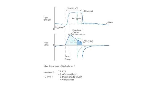
Illustration of pressure and flow
Adapted from Bellani, Giacomo. Mechanical ventilation from pathophysiology to clinical evidence. Cham, Switzerland: Springer, 2022.
Illustration of pressure and flow
Adapted from Bellani, Giacomo. Mechanical ventilation from pathophysiology to clinical evidence. Cham, Switzerland: Springer, 2022.

NIV-ST

In S/T mode, the clinician sets the inspiratory pressure (∆Pinsp) and expiratory pressure (PEEP), respiratory rate, and inspiratory time. The patient may initiate breaths that are supported to the ∆Pinsp level, as in the NIV mode, but if the patient fails to make an inspiratory effort within a set interval (that is defined by the set respiratory rate), the machine triggers inspiration to the set ∆Pinsp level. ∆Pinsp then cycles to PEEP based on the inspiratory time period.

NIV-ST with the backup rate is useful in the case of apnea or periodic breathing (Teaching Pearls in Noninvasive Mechanical Ventilation: Key Practical Insights7​).

Illustration of NIV-ST

Pressure-support mode versus CPAP/BPAP

The NIV and NIV-ST modes are types of noninvasive positive pressure ventilation. Pressure support is generally considered to be the same as CPAP/BIPAP, except that pressure support is delivered by a ventilator and BIPAP through a noninvasive ventilator.

In NIV and NIV-ST, the level of pressure support is applied as pressure above baseline PEEP. However, the approach is different with bilevel ventilators where IPAP (inspiratory positive airway pressure) and EPAP (expiratory positive airway pressure) are set. In this configuration, the difference between IPAP and EPAP is the level of pressure support.
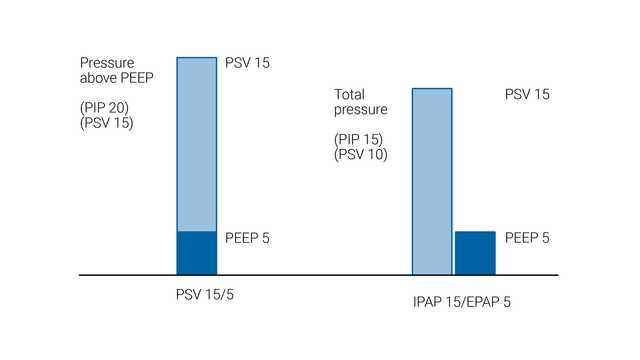
Comparison of PSV and IPAP
Comparison of pressure support ventilation (PSV), such as with critical care ventilators, and inspiratory positive airway pressure (IPAP) with a bilevel ventilator. Note that IPAP is the peak inspiratory pressure (PIP) and includes the expiratory positive airway pressure (EPAP), whereas in PSV, pressure support is provided on top of the positive end-expiratory pressure (PEEP).

When to consider noninvasive ventilation?

In order to minimize the risk of failure or complications, every patient should be properly assessed for suitability to receive NIV safely.

Indications for NIV use include patients who have:

  • Dyspnea
  • Tachypnea
  • Accessory respiratory muscle use
  • Paradoxical abdominal “belly” breathing
  • PaCO2 > 45 mmHg and pH < 7.35 (Ozyilmaz E, Ugurlu AO, Nava S. Timing of noninvasive ventilation failure: causes, risk factors, and potential remedies. BMC Pulm Med. 2014;14:19. Published 2014 Feb 13. doi:10.1186/1471-2466-14-198​,Scala R, Pisani L. Noninvasive ventilation in acute respiratory failure: which recipe for success?. Eur Respir Rev. 2018;27(149):180029. Published 2018 Jul 11. doi:10.1183/16000617.0029-20189​)

Patients should be closely monitored during the first 24 hours after initiating NIV, as this is the period with the highest rate of treatment failure. Although data points at presentation such as a high RR (respiratory rate), low arterial pH values or low PaO2/FiO2 can help predict failure, the most robust predictor of treatment failure during this period is failing to show an improvement in these parameters at 1–2 h after initiating NIV treatment (Scala R, Pisani L. Noninvasive ventilation in acute respiratory failure: which recipe for success?. Eur Respir Rev. 2018;27(149):180029. Published 2018 Jul 11. doi:10.1183/16000617.0029-20189​).

Indications and recommendations for NPPV

Clinical indication (Rochwerg B, Brochard L, Elliott MW, et al. Official ERS/ATS clinical practice guidelines: noninvasive ventilation for acute respiratory failure. Eur Respir J. 2017;50(2):1602426. Published 2017 Aug 31. doi:10.1183/13993003.02426-20161​, Davidson AC, Banham S, Elliott M, et al. BTS/ICS guideline for the ventilatory management of acute hypercapnic respiratory failure in adults [published correction appears in Thorax. 2017 Jun;72 (6):588]. Thorax. 2016;71 Suppl 2:ii1-ii35. doi:10.1136/thoraxjnl-2015-2082096​)

Certainty of evidence

Recommendation

Hypercapnia with COPD exacerbation

High

Strong recommendation for

Cardiogenic pulmonary edema (CPE)

Moderate

Strong recommendation for

Immunocompromised

Moderate

Conditional recommendation for

Post-operative patients

Moderate

Conditional recommendation for

Palliative care

Moderate

Conditional recommendation for

Trauma

Moderate

Conditional recommendation for

Weaning in hypercapnic patients

Moderate

Conditional recommendation for

Post-extubation respiratory failure

Low

Conditional recommendation against

Obesity hypoventilation syndrome (OHS)

Low

Conditional recommendation for

Neuromuscular disease and chest wall disease

Low

Conditional recommendation for

Prevention of hypercapnia in COPD exacerbation

Low

Conditional recommendation against

Post-extubation in high-risk patients (prophylaxis)

Low

Conditional recommendation for

De novo respiratory failure

No certain evidence

No recommendation made

Acute asthma exacerbation

No certain evidence

No recommendation made

Contraindications to noninvasive ventilation

Absolute:

  • The need for emergent intubation (i.e., cardiac or respiratory arrest, severe respiratory distress, unstable cardiac arrhythmia)

Relative:

  • Non-respiratory organ failure that is acutely life-threatening
  • Severe encephalopathy (i.e., Glasgow coma score < 10)
  • Severe upper gastrointestinal bleeding
  • Hemodynamic instability
  • Facial or neurological surgery, trauma, or deformity
  • Significant airway obstruction (i.e., laryngeal mass or tracheal tumor)
  • Inability to cooperate, protect airway, or clear secretions (i.e., patients at high risk of aspiration)
  • Anticipated prolonged duration of mechanical ventilation (i.e., ≥ 4 to 7 days)
  • Recent esophageal or gastric anastomosis (GCS: Glasgow Coma Score and esophageal or gastric distension from air may increase the risk of anastomotic dehiscenceA​)
  • Multiple contraindications
  • Insufficient staffing support

(Evans TW. International Consensus Conferences in Intensive Care Medicine: non-invasive positive pressure ventilation in acute respiratory failure. Organised jointly by the American Thoracic Society, the European Respiratory Society, the European Society of Intensive Care Medicine, and the Société de Réanimation de Langue Française, and approved by the ATS Board of Directors, December 2000. Intensive Care Med. 2001;27(1):166-178. doi:10.1007/s00134000072110​)

Illustration for the chapter "What is the clinical relevance of NIV?"

What is the clinical relevance of NIV? Physiologic effects of noninvasive ventilation

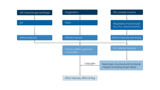
The primary desired effect of NIV is to maintain adequate levels of PO2 and PCO2 in the arterial blood while also unloading the inspiratory muscles.

The physiologic effects of noninvasive ventilation are the following: 

  • Augments minute ventilation
  • Unloads ventilatory muscles
  • Resets the ventilatory control system
  • Improves alveolar recruitment and gas exchange
  • Maintains upper-airway patency
  • Reduces triggering loads from Auto-PEEP
  • Reduces the risk of ventilator-induced lung injury

(MacIntyre NR. Physiologic Effects of Noninvasive Ventilation. Respir Care. 2019;64(6):617-628. doi:10.4187/respcare.0663511​)

Illustration of the physiologic effects of NIV
Adapted from MacIntyre, Neil R. Physiologic effects of noninvasive ventilation. Respiratory care 64.6 (2019): 617-628
Illustration of the physiologic effects of NIV
Adapted from MacIntyre, Neil R. Physiologic effects of noninvasive ventilation. Respiratory care 64.6 (2019): 617-628
Illustration for the chapter "How to start a patient on NIV?"

How to start a patient on NIV?

  • Education: Plan adequate training for all staff with a calibrated protocol
  • Environment: Choose an appropriate setting for starting NIV according to the severity of ARF
  • Indication: Select patients according to the team's experience, location, availability of intubation, do-not-intubate status, and likelihood of success
  • Information: Explain the technique to competent patients to improve their compliance
  • Equipment: Choose the interface(s) that best fits the facial anatomy; also consider rotating different interfaces to enhance comfort. Choose a ventilator with good air-leak compensation that displays flow/pressure/volume curves
  • Starting ventilation: Choose a pressure mode (i.e., pressure support) with PEEP. Start with low pressures, then increase gradually depending on comfort. Set adequate FiO2 and essential alarms. Tighten the straps of the interface enough to avoid leaks, without making them too tight
  • Monitoring ventilation: Check clinical status, monitor SpO2, measure blood gases periodically. Reset the ventilator according to patient–ventilator synchrony, comfort, and leaks. Prevent skin lesions (i.e., protective devices, rotating interfaces). Consider humidification. Carefully consider sedation. Consider management of secretions, if required.

(Non-invasive ventilation and weaning: principles and practice12​)

Conditions for initiating NIV

  • Choose a location with appropriate monitoring based on the severity of the patient’s condition. At a minimum, continuous pulse oximetry should be provided.
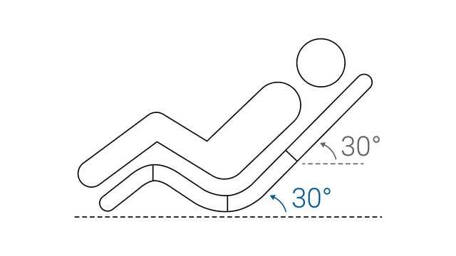
  • Optimizing the patient’s position also plays a key role in ensuring comfort during NIV (Non–Invasive Ventilation Guidelines for Adult Patients With Acute Respiratory Failure: A Clinical Practice Guideline.13​). A sitting or semi-recumbent position is suggested during NIV to ensure a high level of comfort to patients. A side-lying position may help to remove pressure from a pendulous abdomen, as in case of pregnancy or obesity (Non–Invasive Ventilation Guidelines for Adult Patients With Acute Respiratory Failure: A Clinical Practice Guideline.13​). Recently, the use of the prone positioning has been introduced in patients with ARF, particularly those with COVID-19 disease (Ehrmann S, Li J, Ibarra-Estrada M, et al. Awake prone positioning for COVID-19 acute hypoxaemic respiratory failure: a randomised, controlled, multinational, open-label meta-trial. Lancet Respir Med. 2021;9(12):1387-1395. doi:10.1016/S2213-2600(21)00356-814​). The analysis of this rescue therapy is better explained in the last paragraph on the COVID-19 pandemic.
  • Select a ventilator and an appropriately sized interface, and ensure the interface is compatible with the type of ventilator to be used.
Illustration of a patient in semi-recumbent position
Illustration for the chapter "How to choose the right NIV interface?"

How to choose the right interface for NIV?

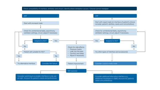
Select the mask designed for use with a critical care ventilator (without entrainment valves or leak port). 

The wrong size of mask, incorrect positioning, or an insufficient mask seal may all lead to the patient discomfort and pressure injuries. It is important to choose an interface that is the correct size for the individual patient, evaluate the mask fit and placement on the face, and reposition the mask as needed to minimize leaks. 

Good to know: NIV interfaces with entrainment valves are designed for noninvasive ventilators that use a single-limb circuit. The entrainment valve is required to prevent asphyxia if the ventilator fails or the tubing becomes disconnected. Masks with leak ports should only be used with single-limb circuit ventilators and should not be used with Hamilton Medical ventilators.

(Simonds, A. K. (Ed.). (2015). ERS practical handbook of noninvasive ventilation. European Respiratory Society.15)

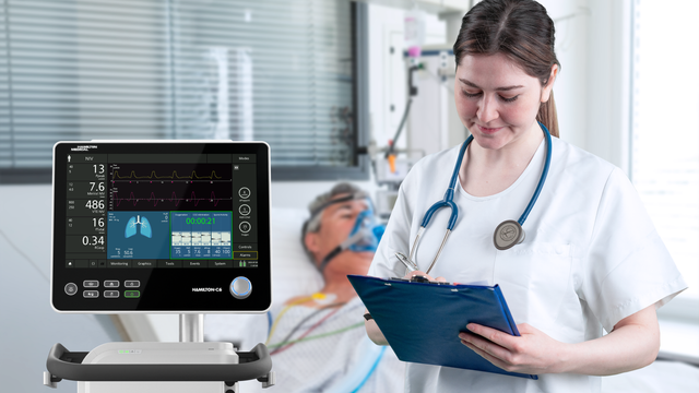
Nurse with a NIV patient and the HAMILTON-C6 ventilator
Illustration of a compatibilit tree for NIV masks
Adapted from Simonds, Anita K., ed. ERS practical handbook of noninvasive ventilation. European Respiratory Society, 2015.
Illustration of a compatibilit tree for NIV masks
Adapted from Simonds, Anita K., ed. ERS practical handbook of noninvasive ventilation. European Respiratory Society, 2015.
Illustration for the chapter "How to set up NIV?"

How to set up NIV?

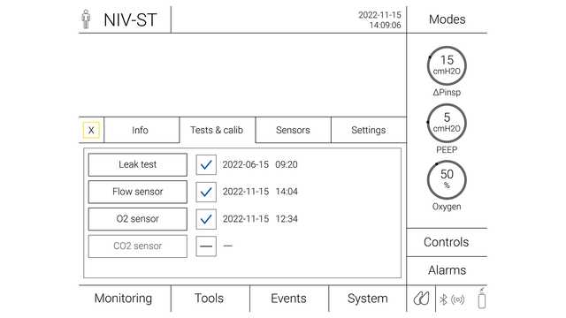
  • Turn on the ventilator and humidifier, then run the calibration and tightness test
  • Connect the interface to the circuit
  • Explain the procedure and reason for therapy to the patient, and answer any questions about NIV before placing the mask on the patient
Illustration of a ventilator screen with the test and calibration for NIV-ST

Humidification during NIV

Recommendations (Holanda MA, Reis RC, Winkeler GF, Fortaleza SC, Lima JW, Pereira ED. Influence of total face, facial and nasal masks on short-term adverse effects during noninvasive ventilation. J Bras Pneumol. 2009;35(2):164-173. doi:10.1590/s1806-3713200900020001016​, American Association for Respiratory Care, Restrepo RD, Walsh BK. Humidification during invasive and noninvasive mechanical ventilation: 2012. Respir Care. 2012;57(5):782-788. doi:10.4187/respcare.0176617​):

  • All NIV circuits are to be actively humidified
  • HMEs (heat moisture exchanger) are not recommended for NIV
  • Gas temperatures during NIV are to be based on patient comfort
  • Humidified gas, heated to a temperature that is comfortable for the patient (usually about 30°C), should be the standard when using NIV

Patient agreement and motivation

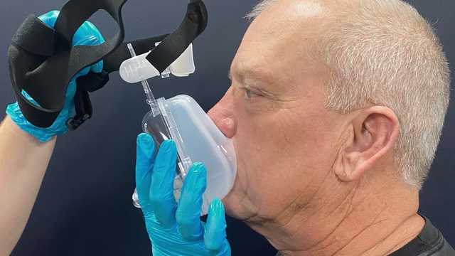
Dyspnea can cause feelings of anxiety and fear. For this reason, the healthcare professional or the patient should hold the mask in place when applying it for the first time.

This way the mask can be removed quickly if the patient begins to panic or needs to communicate. A strategy of starting with low pressures can help patients adjust to NIV more readily.

Patient and NIV mask
Illustration for the chapter "How to adjust NIV settings?"

How to adjust settings? Initial ventilator settings for NIV

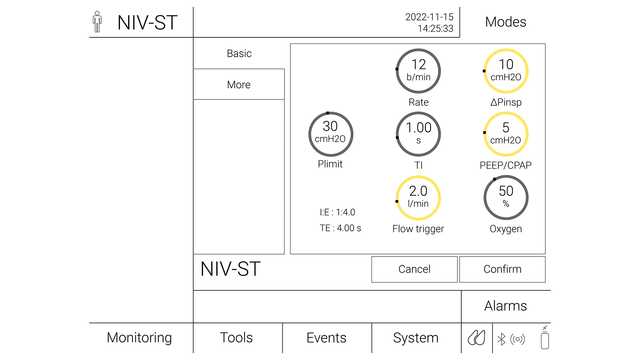
  • Ventilating pressures should be set as low as possible to start with, especially if the patient is unfamiliar with the sensation of positive pressure ventilation (PEEP 4–6 cmH2O, ∆Pinsp 6–10 cmH2O)
  • Ventilating pressures can then be adjusted in small increments over 1 to 2 minutes until exhaled VT is 6 to 8 ml/kg predicted body weight, or respiratory distress improves
  • Adjust FiO2 to keep SpO2 in the desired range (88%–95%)
  • Flow trigger: Set trigger sensitive enough to allow easy triggering without auto-triggering, even in the presence of leaks
  • Peak airway pressures greater than 30 cmH2O are rarely required and are best avoided. Airway pressure settings greater than 30 cmH2O can force air through the esophagus into the stomach (Plimit should not exceed 30 cmH2O or PEEP 8 cmH2O without expert reviewC​)
  • Set the backup rate to 12–16 bpm (2 bpm below resting respiratory rate)
  • Set an appropriate inspiratory time and I:E ratio for the patient’s presenting condition (Ti and I:E ratio (1:2–1:3 (COPD) or 1:1 (NMD/OHS)) set by a competent practitionerD​)
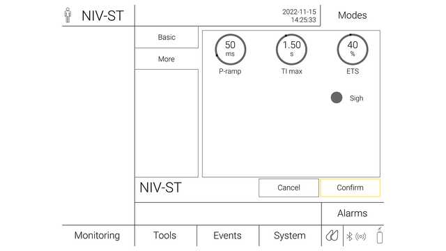
  • ETS: 40%–50%
  • P-ramp (pressure ramp): Should be tailored for a faster slope while avoiding excessive peak flow
  • Maximum inspiratory time is set 0.25 s above the actual inspiratory time; if you are unsure start at 1.5 s

(Developed from BTS/ICS guidelines (https://www.brit-thoracic.org.uk/document-library/clinical-information/acute-hypercapnic-respiratory-failure/bts-guidelines-for-ventilatory-management-of-ahrf/)B​)

Illustration of a ventilator screen settings for NIV-ST
Illustration of a ventilator screen settings for ETS and Pramp for NIV-ST

Alarm setup for NIV

Emergency alarms are important for recognizing a deterioration in the patient's condition and alert staff in the following situations:
  • Apnea: when the patient stops breathing
  • High respiratory rate: when the patient’s respiratory rate goes above set value
  • Low respiratory rate: when the patient’s respiratory rate goes below a set value
  • High tidal volume: when the patient’s tidal volume goes above a target value
  • Low tidal volume: when the patient’s tidal volume goes below a target value
Illustration for the chapter "How should I monitor patients on NIV?"

How should I monitor patients on NIV?

(Developed from BTS/ICS guidelines (https://www.brit-thoracic.org.uk/document-library/clinical-information/acute-hypercapnic-respiratory-failure/bts-guidelines-for-ventilatory-management-of-ahrf/)B​)

  • Reassess frequently for tolerance and efficacy of NIV (at least every 30 min) for the first 1 to 2 h
  • Continuous cardiac and SpO2 monitoring for at least the first 12 hours. Ensure PaCO2, PaO2, and SpO2 parameters are set
  • Alter NIV settings: If PaCO2 remains high, increase tidal volume (Vt) by increasing ∆Pinsp. If the patient remains hypoxic, increase PEEP or FiO2 (remember you may need to increase ∆Pinsp to maintain Vt between 6–8 ml/kg or signs of respiratory distress improve), update NIV prescription, and repeat ABG (arterial blood gases) one hour after any settings are changed (Increase ∆Pinsp over 10-30 minutes. Ppeak should not exceed 30 cmH2O or PEEP 8 cmH2O without expert reviewE​)
  • Check the patient's tolerance with the initial settings, and assess for dyspnea and asynchrony
  • Monitor the patient for risk of possible NIV failure
  • Use prespecified criteria for escalation
  • Do not delay an indicated intubation

Monitoring failure during noninvasive ventilation

Failure of NIV has usually been defined as a need for intubation due to a lack of improvement in arterial blood gases and clinical parameters, or death (Teaching Pearls in Noninvasive Mechanical Ventilation: Key Practical Insights7​). It is very important to identify patients who are at risk of failing NIV, because an inappropriate delay in intubation may cause an increase in morbidity and mortality.

Success predictors:

  • Increase in arterial oxygenation
  • Decrease in respiratory rate
  • Decrease in dyspnea
  • Significant increase in PaO2/FiO2 (Partial pressure of oxygen/Fraction of inspired oxygen)

Failure predictors:

  • Stable or decrease in arterial oxygenation
  • Stable or rise in respiratory rate
  • Stable or increase in dyspnea
  • Stable or decrease in PaO2/FiO2
  • Stable or increase in OI (oxygen index)
  • Presence of contraindication(s) to NIV

In a large prospective cohort study, the HACOR scale predicted NIV failure after 1 h of treatment with high specificity (90%) and good sensitivity (72%) (Duan J, Han X, Bai L, Zhou L, Huang S. Assessment of heart rate, acidosis, consciousness, oxygenation, and respiratory rate to predict noninvasive ventilation failure in hypoxemic patients. Intensive Care Med. 2017;43(2):192-199. doi:10.1007/s00134-016-4601-318​)

How to monitor patient-ventilator synchrony?

Patient-ventilator synchronization is an important issue that can influence the efficacy and success of NIV.

The most common phenomenon is ineffective triggering (patient effort is not recognized by the ventilator; may be secondary to high AutoPEEP or inappropriate inspiratory trigger sensitivity), followed by auto-triggering (delivery of preset pressure in the absence of patient effort) and double triggering (consecutive delivery of two preset pressure support events within an interval of less than half the mean inspiratory time due to the patient’s continued effort) (de Wit M, Miller KB, Green DA, Ostman HE, Gennings C, Epstein SK. Ineffective triggering predicts increased duration of mechanical ventilation. Crit Care Med. 2009;37(10):2740-2745. doi:10.1097/ccm.0b013e3181a98a0519​,Epstein SK. How often does patient-ventilator asynchrony occur and what are the consequences?. Respir Care. 2011;56(1):25-38. doi:10.4187/respcare.0100920​,Chao DC, Scheinhorn DJ, Stearn-Hassenpflug M. Patient-ventilator trigger asynchrony in prolonged mechanical ventilation. Chest. 1997;112(6):1592-1599. doi:10.1378/chest.112.6.159221​,Thille AW, Lyazidi A, Richard JC, Galia F, Brochard L. A bench study of intensive-care-unit ventilators: new versus old and turbine-based versus compressed gas-based ventilators. Intensive Care Med. 2009;35(8):1368-1376. doi:10.1007/s00134-009-1467-722​)

Synchrony between the patient and ventilator should be checked frequently. Asynchronies can be detected by observing the patient and asking them simple questions. The most practical method should be analysis of the pressure and flow waveforms (Ergan B, Nasiłowski J, Winck JC. How should we monitor patients with acute respiratory failure treated with noninvasive ventilation?. Eur Respir Rev. 2018;27(148):170101. Published 2018 Apr 13. doi:10.1183/16000617.0101-201723​).

Hamilton Medical ventilators offer a functionality for leak compensation, IntelliTrig, during the full breath cycle to increase patient-ventilator synchrony and reduce the risk of auto-triggering. Using IntelliTrig, the ventilator identifies the leak by measuring the flow at the airway opening, and uses this data to automatically adjust the gas delivery while remaining responsive to the set inspiratory and expiratory trigger sensitivity.

Hamilton Medical ventilators also have the optional feature, IntelliSync+, which continuously analyzes waveform shapes and is able to detect patient efforts immediately, then initiate inspiration or expiration in real-time (IntelliSync+ is available as an optional feature on the HAMILTON-C6 and HAMILTON-G5 mechanical ventilators, and is standard on the HAMILTON-S1.F​). For maximum flexibility, IntelliSync+ can be activated for either the inspiratory trigger or the expiratory trigger, or both.

Noninvasive ventilation on Hamilton Medical ventilators

Together with our HAMILTON-C1, the compact solution for noninvasive ventilationall Hamilton Medical ventilators offer the NIV option (it is available on the HAMILTON‑C6, the HAMILTON‑C3, and on the HAMILTON‑C1/T1/MR1).
Электронная книга «Неинвазивная вентиляция с положительным давлением (NIV)»

Бесплатная электронная книга

Полезно знать! Основы неинвазивной вентиляции с положительным давлением

Ознакомьтесь с описанием преимуществ и клинической значимости неинвазивной вентиляции, а также практическую информацию о выборе правильного интерфейса, настройке параметров и мониторинге пациентов.

Сноски

  • A. GCS: Glasgow Coma Score and esophageal or gastric distension from air may increase the risk of anastomotic dehiscence
  • B. Developed from BTS/ICS guidelines (https://www.brit-thoracic.org.uk/document-library/clinical-information/acute-hypercapnic-respiratory-failure/bts-guidelines-for-ventilatory-management-of-ahrf/)
  • C. Plimit should not exceed 30 cmH2O or PEEP 8 cmH2O without expert review
  • D. Ti and I:E ratio (1:2–1:3 (COPD) or 1:1 (NMD/OHS)) set by a competent practitioner
  • E. Increase ∆Pinsp over 10-30 minutes. Ppeak should not exceed 30 cmH2O or PEEP 8 cmH2O without expert review
  • F. Функция IntelliSync+ доступна как опция в аппаратах ИВЛ HAMILTON-C6 и HAMILTON-G5, а также входит в стандартную комплектацию аппарата ИВЛ HAMILTON-S1.

Список литературы

  1. 1. Rochwerg B, Brochard L, Elliott MW, et al. Official ERS/ATS clinical practice guidelines: noninvasive ventilation for acute respiratory failure. Eur Respir J. 2017;50(2):1602426. Published 2017 Aug 31. doi:10.1183/13993003.02426-2016
  2. 2. Gong Y, Sankari A. Noninvasive Ventilation. In: StatPearls. Treasure Island (FL): StatPearls Publishing; December 11, 2022.
  3. 3. Egan's fundamentals of respiratory care e-book
  4. 4. Rochwerg B, Einav S, Chaudhuri D, et al. The role for high flow nasal cannula as a respiratory support strategy in adults: a clinical practice guideline. Intensive Care Med. 2020;46(12):2226-2237. doi:10.1007/s00134-020-06312-y
  5. 5. Bello G, De Santis P, Antonelli M. Non-invasive ventilation in cardiogenic pulmonary edema. Ann Transl Med. 2018;6(18):355. doi:10.21037/atm.2018.04.39
  6. 6. Davidson AC, Banham S, Elliott M, et al. BTS/ICS guideline for the ventilatory management of acute hypercapnic respiratory failure in adults [published correction appears in Thorax. 2017 Jun;72 (6):588]. Thorax. 2016;71 Suppl 2:ii1-ii35. doi:10.1136/thoraxjnl-2015-208209
  7. 7. Esquinas, A. M. (Ed.). (2022). Teaching Pearls in Noninvasive Mechanical Ventilation: Key Practical Insights. Springer Nature.
  8. 8. Ozyilmaz E, Ugurlu AO, Nava S. Timing of noninvasive ventilation failure: causes, risk factors, and potential remedies. BMC Pulm Med. 2014;14:19. Published 2014 Feb 13. doi:10.1186/1471-2466-14-19
  9. 9. Scala R, Pisani L. Noninvasive ventilation in acute respiratory failure: which recipe for success?. Eur Respir Rev. 2018;27(149):180029. Published 2018 Jul 11. doi:10.1183/16000617.0029-2018
  10. 10. Evans TW. International Consensus Conferences in Intensive Care Medicine: non-invasive positive pressure ventilation in acute respiratory failure. Organised jointly by the American Thoracic Society, the European Respiratory Society, the European Society of Intensive Care Medicine, and the Société de Réanimation de Langue Française, and approved by the ATS Board of Directors, December 2000. Intensive Care Med. 2001;27(1):166-178. doi:10.1007/s001340000721
  11. 11. MacIntyre NR. Physiologic Effects of Noninvasive Ventilation. Respir Care. 2019;64(6):617-628. doi:10.4187/respcare.06635
  12. 12. Elliott, M., Nava, S., & Schönhofer, B. (Eds.). (2018). Non-invasive ventilation and weaning: principles and practice. CRC Press.
  13. 13. Sanchez D, Smith G, Piper A, Rolls K. Non–Invasive Ventilation Guidelines for Adult Patients With Acute Respiratory Failure: A Clinical Practice Guideline. Agency for clinical innovation NSW government Version 1, Chatswood NSW, ISBN 978-1-74187-954-4 (2014).
  14. 14. Ehrmann S, Li J, Ibarra-Estrada M, et al. Awake prone positioning for COVID-19 acute hypoxaemic respiratory failure: a randomised, controlled, multinational, open-label meta-trial. Lancet Respir Med. 2021;9(12):1387-1395. doi:10.1016/S2213-2600(21)00356-8
  15. 15. Simonds, A. K. (Ed.). (2015). ERS practical handbook of noninvasive ventilation. European Respiratory Society.
  16. 16. Holanda MA, Reis RC, Winkeler GF, Fortaleza SC, Lima JW, Pereira ED. Influence of total face, facial and nasal masks on short-term adverse effects during noninvasive ventilation. J Bras Pneumol. 2009;35(2):164-173. doi:10.1590/s1806-37132009000200010
  17. 17. American Association for Respiratory Care, Restrepo RD, Walsh BK. Humidification during invasive and noninvasive mechanical ventilation: 2012. Respir Care. 2012;57(5):782-788. doi:10.4187/respcare.01766
  18. 18. Duan J, Han X, Bai L, Zhou L, Huang S. Assessment of heart rate, acidosis, consciousness, oxygenation, and respiratory rate to predict noninvasive ventilation failure in hypoxemic patients. Intensive Care Med. 2017;43(2):192-199. doi:10.1007/s00134-016-4601-3
  19. 19. de Wit M, Miller KB, Green DA, Ostman HE, Gennings C, Epstein SK. Ineffective triggering predicts increased duration of mechanical ventilation. Crit Care Med. 2009;37(10):2740-2745. doi:10.1097/ccm.0b013e3181a98a05
  20. 20. Epstein SK. How often does patient-ventilator asynchrony occur and what are the consequences?. Respir Care. 2011;56(1):25-38. doi:10.4187/respcare.01009
  21. 21. Chao DC, Scheinhorn DJ, Stearn-Hassenpflug M. Patient-ventilator trigger asynchrony in prolonged mechanical ventilation. Chest. 1997;112(6):1592-1599. doi:10.1378/chest.112.6.1592
  22. 22. Thille AW, Lyazidi A, Richard JC, Galia F, Brochard L. A bench study of intensive-care-unit ventilators: new versus old and turbine-based versus compressed gas-based ventilators. Intensive Care Med. 2009;35(8):1368-1376. doi:10.1007/s00134-009-1467-7
  23. 23. Ergan B, Nasiłowski J, Winck JC. How should we monitor patients with acute respiratory failure treated with noninvasive ventilation?. Eur Respir Rev. 2018;27(148):170101. Published 2018 Apr 13. doi:10.1183/16000617.0101-2017

Official ERS/ATS clinical practice guidelines: noninvasive ventilation for acute respiratory failure.

Rochwerg B, Brochard L, Elliott MW, et al. Official ERS/ATS clinical practice guidelines: noninvasive ventilation for acute respiratory failure. Eur Respir J. 2017;50(2):1602426. Published 2017 Aug 31. doi:10.1183/13993003.02426-2016

Noninvasive mechanical ventilation (NIV) is widely used in the acute care setting for acute respiratory failure (ARF) across a variety of aetiologies. This document provides European Respiratory Society/American Thoracic Society recommendations for the clinical application of NIV based on the most current literature.The guideline committee was composed of clinicians, methodologists and experts in the field of NIV. The committee developed recommendations based on the GRADE (Grading, Recommendation, Assessment, Development and Evaluation) methodology for each actionable question. The GRADE Evidence to Decision framework in the guideline development tool was used to generate recommendations. A number of topics were addressed using technical summaries without recommendations and these are discussed in the supplementary material.This guideline committee developed recommendations for 11 actionable questions in a PICO (population-intervention-comparison-outcome) format, all addressing the use of NIV for various aetiologies of ARF. The specific conditions where recommendations were made include exacerbation of chronic obstructive pulmonary disease, cardiogenic pulmonary oedema, de novo hypoxaemic respiratory failure, immunocompromised patients, chest trauma, palliation, post-operative care, weaning and post-extubation.This document summarises the current state of knowledge regarding the role of NIV in ARF. Evidence-based recommendations provide guidance to relevant stakeholders.

Noninvasive Ventilation

Gong Y, Sankari A. Noninvasive Ventilation. In: StatPearls. Treasure Island (FL): StatPearls Publishing; December 11, 2022.

Non-invasive ventilation (NIV) was first reported in the mid-eighteen century by a Scottish physician, John Dalziel. In 1864, Alfred F. Jones' patented the first American tank respirator in the iron lung, known as non-invasive negative pressure ventilation. In 1938 a new form of NIV was described by Barach et al. as a treatment for pulmonary edema. However, Oertel described the use of intermittent positive pressure (NPPV) earlier by Oertel (1878).  During the polio epidemic and due to very high mortality (more than 80%), innovation was sparked by physicians such as Bjorn Ibsen, an anesthesiologist from Copenhagen, Denmark, who applied positive pressure ventilation in 1952 via trach but required manual delivery. The approach dropped the mortality by more than half (to nearly 40%); however, the delivery of pressure was a logistical problem, as there were no positive pressure ventilators, and patients needed to be bagged by hand. Over the past century, positive pressure ventilation (NPPV) has been dramatically improved and used to treat respiratory failure from multiple etiologies. It has been proven effective in preventing intubation compared to standard oxygen therapy in the acute setting. NPPV encompasses several methods of respiratory support, the most common being Bilevel Positive Airway Pressure (BPAP). The latest American Thoracic Society/European Respiratory Journal guidelines support the use of NPPV in acute exacerbation of chronic obstructive pulmonary disease (AECOPD) and acute respiratory failure secondary to cardiogenic pulmonary edema, where evidence and level of recommendation are the strongest. However, there is a body of evidence and conditional recommendations that NPPV is effective in other settings of acute respiratory failure, such as post-operative and chest trauma. In addition, several studies support the use of NPPV in various chronic respiratory diseases.

Egan's fundamentals of respiratory care e-book

The role for high flow nasal cannula as a respiratory support strategy in adults: a clinical practice guideline.

Rochwerg B, Einav S, Chaudhuri D, et al. The role for high flow nasal cannula as a respiratory support strategy in adults: a clinical practice guideline. Intensive Care Med. 2020;46(12):2226-2237. doi:10.1007/s00134-020-06312-y


PURPOSE

High flow nasal cannula (HFNC) is a relatively recent respiratory support technique which delivers high flow, heated and humidified controlled concentration of oxygen via the nasal route. Recently, its use has increased for a variety of clinical indications. To guide clinical practice, we developed evidence-based recommendations regarding use of HFNC in various clinical settings.

METHODS

We formed a guideline panel composed of clinicians, methodologists and experts in respiratory medicine. Using GRADE, the panel developed recommendations for four actionable questions.

RESULTS

The guideline panel made a strong recommendation for HFNC in hypoxemic respiratory failure compared to conventional oxygen therapy (COT) (moderate certainty), a conditional recommendation for HFNC following extubation (moderate certainty), no recommendation regarding HFNC in the peri-intubation period (moderate certainty), and a conditional recommendation for postoperative HFNC in high risk and/or obese patients following cardiac or thoracic surgery (moderate certainty).

CONCLUSIONS

This clinical practice guideline synthesizes current best-evidence into four recommendations for HFNC use in patients with hypoxemic respiratory failure, following extubation, in the peri-intubation period, and postoperatively for bedside clinicians.

Non-invasive ventilation in cardiogenic pulmonary edema.

Bello G, De Santis P, Antonelli M. Non-invasive ventilation in cardiogenic pulmonary edema. Ann Transl Med. 2018;6(18):355. doi:10.21037/atm.2018.04.39

Cardiogenic pulmonary edema (CPE) is among the most common causes of acute respiratory failure (ARF) in the acute care setting and often requires ventilatory assistance. In patients with ARF due to CPE, use of non-invasive positive airway pressure can decrease the systemic venous return and the left ventricular (LV) afterload, thus reducing LV filling pressure and limiting pulmonary edema. In these patients, either non-invasive ventilation (NIV) or continuous positive airway pressure (CPAP) can improve vital signs and physiological parameters, decreasing the need for endotracheal intubation (ETI) and hospital mortality when compared to conventional oxygen therapy. Results on the use of NIV or CPAP in patients with CPE prior to hospitalization are not homogeneous among studies, hampering any conclusive recommendation regarding their role in the pre-hospital setting.

BTS/ICS guideline for the ventilatory management of acute hypercapnic respiratory failure in adults.

Davidson AC, Banham S, Elliott M, et al. BTS/ICS guideline for the ventilatory management of acute hypercapnic respiratory failure in adults [published correction appears in Thorax. 2017 Jun;72 (6):588]. Thorax. 2016;71 Suppl 2:ii1-ii35. doi:10.1136/thoraxjnl-2015-208209

Teaching Pearls in Noninvasive Mechanical Ventilation: Key Practical Insights

Presents clinical cases Offers a practical guide to evaluating and diagnosing acute respiratory failure for NIV Describe how to evaluate parameter settings, ventilatory modes and interfaces Discusses major hot topics for pneumologists, intensivists/anaesthesiologists

Timing of noninvasive ventilation failure: causes, risk factors, and potential remedies.

Ozyilmaz E, Ugurlu AO, Nava S. Timing of noninvasive ventilation failure: causes, risk factors, and potential remedies. BMC Pulm Med. 2014;14:19. Published 2014 Feb 13. doi:10.1186/1471-2466-14-19


BACKGROUND

Identifying the predictors of noninvasive ventilation (NIV) failure has attracted significant interest because of the strong link between failure and poor outcomes. However, very little attention has been paid to the timing of the failure. This narrative review focuses on the causes of NIV failure and risk factors and potential remedies for NIV failure, based on the timing factor.

RESULTS

The possible causes of immediate failure (within minutes to <1 h) are a weak cough reflex, excessive secretions, hypercapnic encephalopathy, intolerance, agitation, and patient-ventilator asynchrony. The major potential interventions include chest physiotherapeutic techniques, early fiberoptic bronchoscopy, changing ventilator settings, and judicious sedation. The risk factors for early failure (within 1 to 48 h) may differ for hypercapnic and hypoxemic respiratory failure. However, most cases of early failure are due to poor arterial blood gas (ABGs) and an inability to promptly correct them, increased severity of illness, and the persistence of a high respiratory rate. Despite a satisfactory initial response, late failure (48 h after NIV) can occur and may be related to sleep disturbance.

CONCLUSIONS

Every clinician dealing with NIV should be aware of these risk factors and the predicted parameters of NIV failure that may change during the application of NIV. Close monitoring is required to detect early and late signs of deterioration, thereby preventing unavoidable delays in intubation.

Noninvasive ventilation in acute respiratory failure: which recipe for success?

Scala R, Pisani L. Noninvasive ventilation in acute respiratory failure: which recipe for success?. Eur Respir Rev. 2018;27(149):180029. Published 2018 Jul 11. doi:10.1183/16000617.0029-2018

Noninvasive positive-pressure ventilation (NPPV) to treat acute respiratory failure has expanded tremendously over the world in terms of the spectrum of diseases that can be successfully managed, the locations of its application and achievable goals.The turning point for the successful expansion of NPPV is its ability to achieve the same physiological effects as invasive mechanical ventilation with the avoidance of the life-threatening risks correlated with the use of an artificial airway.Cardiorespiratory arrest, extreme psychomotor agitation, severe haemodynamic instability, nonhypercapnic coma and multiple organ failure are absolute contraindications for NPPV. Moreover, pitfalls of NPPV reduce its rate of success; consistently, a clear plan of what to do in case of NPPV failure should be considered, especially for patients managed in unprotected setting. NPPV failure is likely to be reduced by the application of integrated therapeutic tools in selected patients handled by expert teams.In conclusion, NPPV has to be considered as a rational art and not just as an application of science, which requires the ability of clinicians to both choose case-by-case the best "ingredients" for a "successful recipe" (i.e. patient selection, interface, ventilator, interface, etc) and to avoid a delayed intubation if the ventilation attempt fails.

International Consensus Conferences in Intensive Care Medicine: non-invasive positive pressure ventilation in acute respiratory failure. Organised jointly by the American Thoracic Society, the European Respiratory Society, the European Society of Intensive Care Medicine, and the Société de Réanimation de Langue Française, and approved by the ATS Board of Directors, December 2000.

Evans TW. International Consensus Conferences in Intensive Care Medicine: non-invasive positive pressure ventilation in acute respiratory failure. Organised jointly by the American Thoracic Society, the European Respiratory Society, the European Society of Intensive Care Medicine, and the Société de Réanimation de Langue Française, and approved by the ATS Board of Directors, December 2000. Intensive Care Med. 2001;27(1):166-178. doi:10.1007/s001340000721

Physiologic Effects of Noninvasive Ventilation.

MacIntyre NR. Physiologic Effects of Noninvasive Ventilation. Respir Care. 2019;64(6):617-628. doi:10.4187/respcare.06635

Noninvasive ventilation (NIV) has a number of physiologic effects similar to invasive ventilation. The major effects are to augment minute ventilation and reduce muscle loading. These effects, in turn, can have profound effects on the patient's ventilator control system, both acutely and chronically. Because NIV can be supplied with PEEP, the maintenance of alveolar recruitment is also made possible and the triggering load imposed by auto-PEEP can be reduced. NIV (or simply mask CPAP) can maintain upper-airway patency during sleep in patients with obstructive sleep apnea. NIV can have multiple effects on cardiac function. By reducing venous return, it can help in patients with heart failure or fluid overload, but it can compromise cardiac output in others. NIV can also increase right ventricular afterload or function to reduce left ventricular afterload. Potential detrimental physiologic effects of NIV are ventilator-induced lung injury, auto-PEEP development, and discomfort/muscle overload from poor patient-ventilator interactions.

Non-invasive ventilation and weaning: principles and practice

Now in full-colour, this eagerly-anticipated second edition continues to be the most comprehensive resource available on non-invasive ventilation (NIV), both in the hospital and at home. Reflecting a global perspective with expert contributors from more than 15 countries, the book: • provides clinical examples of NIV in practice with insightful vignettes • covers home- and intensive care-based ventilation • details NIV use in acute and chronic respiratory failure, plus paediatric and other specialty applications. Disease-specific sections provide best practice in the science, diagnostics and management of conditions such as COPD, cardiac failure, neuromuscular disease and obesity, while features such as ‘Common Clinical Questions & Answers’, abundant tables and illustrations, chapter summaries and new clinical vignettes showcase the realities of NIV in practice. This is essential reading for pulmonologists, critical care physicians and intensive care medicine specialists.     

Non–Invasive Ventilation Guidelines for Adult Patients With Acute Respiratory Failure: A Clinical Practice Guideline.

This guide supports local health districts and hospitals to develop local procedures and guidelines for non-invasive ventilation (NIV) for critically ill patients with acute respiratory failure. This guide aims to ensure: patients receive appropriate care at the right time and within the right location of the hospital services are safe, effective and sustainable staff receive appropriate education, training and support.

Awake prone positioning for COVID-19 acute hypoxaemic respiratory failure: a randomised, controlled, multinational, open-label meta-trial.

Ehrmann S, Li J, Ibarra-Estrada M, et al. Awake prone positioning for COVID-19 acute hypoxaemic respiratory failure: a randomised, controlled, multinational, open-label meta-trial. Lancet Respir Med. 2021;9(12):1387-1395. doi:10.1016/S2213-2600(21)00356-8


BACKGROUND

Awake prone positioning has been reported to improve oxygenation for patients with COVID-19 in retrospective and observational studies, but whether it improves patient-centred outcomes is unknown. We aimed to evaluate the efficacy of awake prone positioning to prevent intubation or death in patients with severe COVID-19 in a large-scale randomised trial.

METHODS

In this prospective, a priori set up and defined, collaborative meta-trial of six randomised controlled open-label superiority trials, adults who required respiratory support with high-flow nasal cannula for acute hypoxaemic respiratory failure due to COVID-19 were randomly assigned to awake prone positioning or standard care. Hospitals from six countries were involved: Canada, France, Ireland, Mexico, USA, Spain. Patients or their care providers were not masked to allocated treatment. The primary composite outcome was treatment failure, defined as the proportion of patients intubated or dying within 28 days of enrolment. The six trials are registered with ClinicalTrials.gov, NCT04325906, NCT04347941, NCT04358939, NCT04395144, NCT04391140, and NCT04477655.

FINDINGS

Between April 2, 2020 and Jan 26, 2021, 1126 patients were enrolled and randomly assigned to awake prone positioning (n=567) or standard care (n=559). 1121 patients (excluding five who withdrew from the study) were included in the intention-to-treat analysis. Treatment failure occurred in 223 (40%) of 564 patients assigned to awake prone positioning and in 257 (46%) of 557 patients assigned to standard care (relative risk 0·86 [95% CI 0·75-0·98]). The hazard ratio (HR) for intubation was 0·75 (0·62-0·91), and the HR for mortality was 0·87 (0·68-1·11) with awake prone positioning compared with standard care within 28 days of enrolment. The incidence of prespecified adverse events was low and similar in both groups.

INTERPRETATION

Awake prone positioning of patients with hypoxaemic respiratory failure due to COVID-19 reduces the incidence of treatment failure and the need for intubation without any signal of harm. These results support routine awake prone positioning of patients with COVID-19 who require support with high-flow nasal cannula.

FUNDING

Open AI inc, Rice Foundation, Projet Hospitalier de Recherche Clinique Interrégional, Appel d'Offre 2020, Groupement Interrégional de Recherche Clinique et d'Innovation Grand Ouest, Association pour la Promotion à Tours de la Réanimation Médicale, Fond de dotation du CHRU de Tours, Fisher & Paykel Healthcare Ltd.

ERS Practical Handbook of Noninvasive Ventilation

Simonds, A. K. (Ed.). (2015). ERS practical handbook of noninvasive ventilation. European Respiratory Society.

The ERS Practical Handbook of Noninvasive Ventilation provides a concise “why and how to” guide to NIV from the basics of equipment and patient selection to discharge planning and community care.

Editor Anita K. Simonds has brought together leading clinicians and researchers in the field to provide an easy-to-read guide to all aspects of NIV. This Practical Handbook is a valuable reference and training resource for all NIV practitioners.

Influence of total face, facial and nasal masks on short-term adverse effects during noninvasive ventilation.

Holanda MA, Reis RC, Winkeler GF, Fortaleza SC, Lima JW, Pereira ED. Influence of total face, facial and nasal masks on short-term adverse effects during noninvasive ventilation. J Bras Pneumol. 2009;35(2):164-173. doi:10.1590/s1806-37132009000200010


OBJECTIVE

Failure of noninvasive ventilation (NIV) has been associated with short-term adverse effects related to the use of masks. The aim of this study was to compare the incidence, type and intensity of adverse effects, as well as the comfort, of total face masks (TFMs), facial masks (FMs) and nasal masks (NMs) during NIV.

METHODS

This was a randomized crossover trial involving 24 healthy volunteers submitted to six sessions of NIV in bilevel positive airway pressure mode using the TFM, FM and NM masks at low and moderate-to-high pressure levels. A written questionnaire was applied in order to evaluate eleven specific adverse effects related to the use of the masks. Comfort was assessed using a visual analog scale. The CO2 exhaled into the ventilator circuit was measured between the mask and the exhalation port.

RESULTS

The performance of the TFM was similar to that of the NM and FM in terms of comfort scores. Higher pressure levels reduced comfort and increased adverse effects, regardless of the mask type. When the TFM was used, there were fewer air leaks and less pain at the nose bridge, although there was greater oronasal dryness and claustrophobia. Air leaks were most pronounced when the FM was used. The partial pressure of exhaled CO2 entering the ventilator circuit was zero for the TFM.

CONCLUSIONS

The short-term adverse effects caused by NIV interfaces are related to mask type and pressure settings. The TFM is a reliable alternative to the NM and FM. Rebreathing of CO2 from the circuit is less likely to occur when a TFM is used.

Humidification during invasive and noninvasive mechanical ventilation: 2012.

American Association for Respiratory Care, Restrepo RD, Walsh BK. Humidification during invasive and noninvasive mechanical ventilation: 2012. Respir Care. 2012;57(5):782-788. doi:10.4187/respcare.01766

We searched the MEDLINE, CINAHL, and Cochrane Library databases for articles published between January 1990 and December 2011. The update of this clinical practice guideline is based on 184 clinical trials and systematic reviews, and 10 articles investigating humidification during invasive and noninvasive mechanical ventilation. The following recommendations are made following the Grading of Recommendations Assessment, Development, and Evaluation (GRADE) scoring system: 1. Humidification is recommended on every patient receiving invasive mechanical ventilation. 2. Active humidification is suggested for noninvasive mechanical ventilation, as it may improve adherence and comfort. 3. When providing active humidification to patients who are invasively ventilated, it is suggested that the device provide a humidity level between 33 mg H(2)O/L and 44 mg H(2)O/L and gas temperature between 34°C and 41°C at the circuit Y-piece, with a relative humidity of 100%. 4. When providing passive humidification to patients undergoing invasive mechanical ventilation, it is suggested that the HME provide a minimum of 30 mg H(2)O/L. 5. Passive humidification is not recommended for noninvasive mechanical ventilation. 6. When providing humidification to patients with low tidal volumes, such as when lung-protective ventilation strategies are used, HMEs are not recommended because they contribute additional dead space, which can increase the ventilation requirement and P(aCO(2)). 7. It is suggested that HMEs are not used as a prevention strategy for ventilator-associated pneumonia.

Assessment of heart rate, acidosis, consciousness, oxygenation, and respiratory rate to predict noninvasive ventilation failure in hypoxemic patients.

Duan J, Han X, Bai L, Zhou L, Huang S. Assessment of heart rate, acidosis, consciousness, oxygenation, and respiratory rate to predict noninvasive ventilation failure in hypoxemic patients. Intensive Care Med. 2017;43(2):192-199. doi:10.1007/s00134-016-4601-3


PURPOSE

To develop and validate a scale using variables easily obtained at the bedside for prediction of failure of noninvasive ventilation (NIV) in hypoxemic patients.

METHODS

The test cohort comprised 449 patients with hypoxemia who were receiving NIV. This cohort was used to develop a scale that considers heart rate, acidosis, consciousness, oxygenation, and respiratory rate (referred to as the HACOR scale) to predict NIV failure, defined as need for intubation after NIV intervention. The highest possible score was 25 points. To validate the scale, a separate group of 358 hypoxemic patients were enrolled in the validation cohort.

RESULTS

The failure rate of NIV was 47.8 and 39.4% in the test and validation cohorts, respectively. In the test cohort, patients with NIV failure had higher HACOR scores at initiation and after 1, 12, 24, and 48 h of NIV than those with successful NIV. At 1 h of NIV the area under the receiver operating characteristic curve was 0.88, showing good predictive power for NIV failure. Using 5 points as the cutoff value, the sensitivity, specificity, positive predictive value, negative predictive value, and diagnostic accuracy for NIV failure were 72.6, 90.2, 87.2, 78.1, and 81.8%, respectively. These results were confirmed in the validation cohort. Moreover, the diagnostic accuracy for NIV failure exceeded 80% in subgroups classified by diagnosis, age, or disease severity and also at 1, 12, 24, and 48 h of NIV. Among patients with NIV failure with a HACOR score of >5 at 1 h of NIV, hospital mortality was lower in those who received intubation at ≤12 h of NIV than in those intubated later [58/88 (66%) vs. 138/175 (79%); p = 0.03).

CONCLUSIONS

The HACOR scale variables are easily obtained at the bedside. The scale appears to be an effective way of predicting NIV failure in hypoxemic patients. Early intubation in high-risk patients may reduce hospital mortality.

Ineffective triggering predicts increased duration of mechanical ventilation.

de Wit M, Miller KB, Green DA, Ostman HE, Gennings C, Epstein SK. Ineffective triggering predicts increased duration of mechanical ventilation. Crit Care Med. 2009;37(10):2740-2745. doi:10.1097/ccm.0b013e3181a98a05


OBJECTIVES

To determine whether high rates of ineffective triggering within the first 24 hrs of mechanical ventilation (MV) are associated with longer MV duration and shorter ventilator-free survival (VFS).

DESIGN

Prospective cohort study.

SETTING

Medical intensive care unit (ICU) at an academic medical center.

PATIENTS

Sixty patients requiring invasive MV.

INTERVENTIONS

None.

MEASUREMENTS

Patients had pressure-time and flow-time waveforms recorded for 10 mins within the first 24 hrs of MV initiation. Ineffective triggering index (ITI) was calculated by dividing the number of ineffectively triggered breaths by the total number of breaths (triggered and ineffectively triggered). A priori, patients were classified into ITI >or=10% or ITI <10%. Patient demographics, MV reason, codiagnosis of chronic obstructive pulmonary disease (COPD), sedation levels, and ventilator parameters were recorded.

MEASUREMENTS AND MAIN RESULTS

Sixteen of 60 patients had ITI >or=10%. The two groups had similar characteristics, including COPD frequency and ventilation parameters, except that patients with ITI >or=10% were more likely to have pressured triggered breaths (56% vs. 16%, p = .003) and had a higher intrinsic respiratory rate (22 breaths/min vs. 18, p = .03), but the set ventilator rate was the same in both groups (9 breaths/min vs. 9, p = .78). Multivariable analyses adjusting for pressure triggering also demonstrated that ITI >or=10% was an independent predictor of longer MV duration (10 days vs. 4, p = .0004) and shorter VFS (14 days vs. 21, p = .03). Patients with ITI >or=10% had a longer ICU length of stay (8 days vs. 4, p = .01) and hospital length of stay (21 days vs. 8, p = .03). Mortality was the same in the two groups, but patients with ITI >or=10% were less likely to be discharged home (44% vs. 73%, p = .04).

CONCLUSIONS

Ineffective triggering is a common problem early in the course of MV and is associated with increased morbidity, including longer MV duration, shorter VFS, longer length of stay, and lower likelihood of home discharge.

How often does patient-ventilator asynchrony occur and what are the consequences?

Epstein SK. How often does patient-ventilator asynchrony occur and what are the consequences?. Respir Care. 2011;56(1):25-38. doi:10.4187/respcare.01009

Mechanical ventilation can be life-saving for patients with acute respiratory failure. In between the 2 extremes of complete and no ventilatory support, both patient and machine contribute to ventilatory work. Ideally, ventilator gas delivery would perfectly match patient demand. This patient-ventilator interaction depends on how the ventilator responds to patient respiratory effort and, in turn, how the patient responds to the breath delivered by the ventilator. It is now evident that the interaction between patient and ventilator is frequently suboptimal and that patient-ventilator asynchrony is common. Its prevalence depends on numerous factors, including timing and duration of observation, technique used for detection, patient population, type of asynchrony, ventilation mode and settings (eg, trigger, flow, and cycle criteria), and confounding factors (eg, state of wakefulness, sedation). Patient-ventilator asynchrony is associated with adverse effects, including higher/wasted work of breathing, patient discomfort, increased need for sedation, confusion during the weaning process, prolonged mechanical ventilation, longer stay, and possibly higher mortality. Whether asynchrony is a marker of poor prognosis or causes these adverse outcomes remains to be determined.

Patient-ventilator trigger asynchrony in prolonged mechanical ventilation.

Chao DC, Scheinhorn DJ, Stearn-Hassenpflug M. Patient-ventilator trigger asynchrony in prolonged mechanical ventilation. Chest. 1997;112(6):1592-1599. doi:10.1378/chest.112.6.1592


STUDY OBJECTIVE

To investigate patient-ventilator trigger asynchrony (TA), its prevalence, physiologic basis, and clinical implications in patients requiring prolonged mechanical ventilation (PMV).

STUDY DESIGN

Descriptive and prospective cohort study.

SETTING

Barlow Respiratory Hospital (BRH), a regional weaning center.

PATIENTS

Two hundred consecutive ventilator-dependent patients, transferred to BRH over an 18-month period for attempted weaning from PMV.

METHODS AND INTERVENTIONS

Patients were assessed clinically for TA within the first week of hospital admission, or once they were in hemodynamically stable condition, by observation of uncoupling of accessory respiratory muscle efforts and onset of machine breaths. Patients were excluded if they had weaned by the time of assessment or if they never achieved hemodynamic stability. Ventilator mode was patient triggered, flow control, volume cycled, with a tidal volume of 7 to 10 mL/kg. Esophageal pressure (Peso), airway-opening pressure, and airflow were measured in patients with TA who consented to esophageal catheter insertion. Attempts to decrease TA in each patient included application of positive end-expiratory pressure (PEEP) stepwise to 10 cm H2O, flow triggering, and reduction of ventilator support in pressure support (PS) mode. Patients were followed up until hospital discharge, when outcomes were scored as weaned (defined as >7 days of ventilator independence), failed to wean, or died.

RESULTS

Of the 200 patients screened, 26 were excluded and 19 were found to have TA. Patients with TA were older, carried the diagnosis of COPD more frequently, and had more severe hypercapnia than their counterparts without TA. Only 3 of 19 patients (16%), all with intermittent TA, weaned from mechanical ventilation, after 70, 72, and 108 days, respectively. This is in contrast to a weaning success rate of 57%, with a median (range) time to wean of 33 (3 to 182) days in patients without TA. Observation of uncoupling of accessory respiratory muscle movement and onset of machine breaths was accurate in identifying patients with TA, which was confirmed in all seven patients consenting to Peso monitoring. TA appeared to result from high auto-PEEP and severe pump failure. Adjusting trigger sensitivity and application of flow triggering were unsuccessful in eliminating TA; external PEEP improved but rarely led to elimination of TA that was transient in duration. Reduction of ventilator support in PS mode, with resultant increased respiratory pump output and lower tidal volumes, uniformly succeeded in eliminating TA. However, this approach imposed a fatiguing load on the respiratory muscles and was poorly tolerated.

CONCLUSION

TA can be easily identified clinically, and when it occurs in the patient in stable condition with PMV, is associated with poor outcome.

A bench study of intensive-care-unit ventilators: new versus old and turbine-based versus compressed gas-based ventilators.

Thille AW, Lyazidi A, Richard JC, Galia F, Brochard L. A bench study of intensive-care-unit ventilators: new versus old and turbine-based versus compressed gas-based ventilators. Intensive Care Med. 2009;35(8):1368-1376. doi:10.1007/s00134-009-1467-7


OBJECTIVE

To compare 13 commercially available, new-generation, intensive-care-unit (ICU) ventilators in terms of trigger function, pressurization capacity during pressure-support ventilation (PSV), accuracy of pressure measurements, and expiratory resistance.

DESIGN AND SETTING

Bench study at a research laboratory in a university hospital.

METHODS

Four turbine-based ventilators and nine conventional servo-valve compressed-gas ventilators were tested using a two-compartment lung model. Three levels of effort were simulated. Each ventilator was evaluated at four PSV levels (5, 10, 15, and 20 cm H2O), with and without positive end-expiratory pressure (5 cm H2O). Trigger function was assessed as the time from effort onset to detectable pressurization. Pressurization capacity was evaluated using the airway pressure-time product computed as the net area under the pressure-time curve over the first 0.3 s after inspiratory effort onset. Expiratory resistance was evaluated by measuring trapped volume in controlled ventilation.

RESULTS

Significant differences were found across the ventilators, with a range of triggering delays from 42 to 88 ms for all conditions averaged (P < 0.001). Under difficult conditions, the triggering delay was longer than 100 ms and the pressurization was poor for five ventilators at PSV5 and three at PSV10, suggesting an inability to unload patient's effort. On average, turbine-based ventilators performed better than conventional ventilators, which showed no improvement compared to a bench comparison in 2000.

CONCLUSION

Technical performance of trigger function, pressurization capacity, and expiratory resistance differs considerably across new-generation ICU ventilators. ICU ventilators seem to have reached a technical ceiling in recent years, and some ventilators still perform inadequately.

How should we monitor patients with acute respiratory failure treated with noninvasive ventilation?

Ergan B, Nasiłowski J, Winck JC. How should we monitor patients with acute respiratory failure treated with noninvasive ventilation?. Eur Respir Rev. 2018;27(148):170101. Published 2018 Apr 13. doi:10.1183/16000617.0101-2017

Noninvasive ventilation (NIV) is currently one of the most commonly used support methods in hypoxaemic and hypercapnic acute respiratory failure (ARF). With advancing technology and increasing experience, not only are indications for NIV getting broader, but more severe patients are treated with NIV. Depending on disease type and clinical status, NIV can be applied both in the general ward and in high-dependency/intensive care unit settings with different environmental opportunities. However, it is important to remember that patients with ARF are always very fragile with possible high mortality risk. The delay in recognition of unresponsiveness to NIV, progression of respiratory failure or new-onset complications may result in devastating and fatal outcomes. Therefore, it is crucial to understand that timely action taken according to monitoring variables is one of the key elements for NIV success. The purpose of this review is to outline basic and advanced monitoring techniques for NIV during an ARF episode.