Back

Measurement of AutoPEEP and total PEEP

Article

Author: Clinical Experts Group, Hamilton Medical

Date of first publication: 14.07.2017

Last change: 30.09.2020

(Originally published 14.07.2017) Previously: select Exp hold, when flow=0 select Exp hold again to deactivate hold maneuver. SW versions updated.

In the presence of dynamic pulmonary hyperinflation, the average end-expiratory pressure inside the alveoli (i.e., the actual, total PEEP (PEEPtot)) is higher than the PEEP applied by the ventilator (PEEPe). The difference between PEEPtot and PEEPe corresponds with the intrinsic PEEP (PEEPi), and is also known as AutoPEEP (1).

Measurement of AutoPEEP and total PEEP

AutoPEEP and RCexp

AutoPEEP may also be referred to as air-trapping, breath stacking, dynamic hyperinflation, inadvertent PEEP, or occult PEEP.

AutoPEEP is a common phenomenon in mechanically ventilated patients with long expiratory time constants (RCexp), for example patients with chronic obstructive pulmonary disease or acute severe asthma.

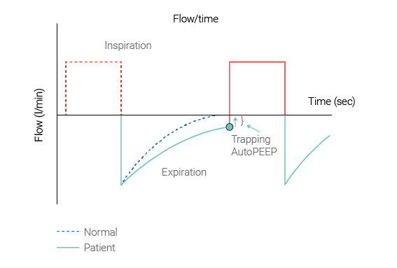
IMPORTANT: The resulting AutoPEEP cannot be seen on the airway pressure curve shown on the ventilator’s screen during normal breath delivery.

(Figure 1 below: Source Garcia Vicente et al. (García Vicente E, Sandoval Almengor JC, Díaz Caballero LA, Salgado Campo JC. Ventilación mecánica invasiva en EPOC y asma [Invasive mechanical ventilation in COPD and asthma]. Med Intensiva. 2011;35(5):288-298. doi:10.1016/j.medin.2010.11.0042​))

Flow-time graph showing AutoPEEP and air trapping
Figure 1: AutoPEEP and air-trapping
Flow-time graph showing AutoPEEP and air trapping
Figure 1: AutoPEEP and air-trapping

Effect of AutoPEEP

AutoPEEP predisposes the patient to increased work of breathing, barotrauma, hemodynamic instability and difficulty in triggering the ventilator. Failure to recognize the hemodynamic consequences of AutoPEEP may lead to inappropriate fluid restriction or unnecessary vasopressor therapy. AutoPEEP can potentially interfere with weaning from mechanical ventilation.

Caregivers should monitor whether AutoPEEP is occurring during ventilation, and set their ventilation control parameters accordingly to avoid the negative consequences of AutoPEEP.

Measuring AutoPEEP

All Hamilton Medical Ventilators have the unique capability of showing AutoPEEP as a monitoring parameter on a breath-by-breath basis. It is calculated using the LSF method applied to the entire breath (Iotti GA, Braschi A, Brunner JX, et al. Respiratory mechanics by least squares fitting in mechanically ventilated patients: applications during paralysis and during pressure support ventilation. Intensive Care Med. 1995;21(5):406-413. doi:10.1007/BF017074093​). However in special circumstances for example when severe dynamic hyperinflation is present, the AutoPEEP calculated by LSF can underestimate the actual AutoPEEP. In these cases it can be obtained by performing an expiratory hold maneuver.

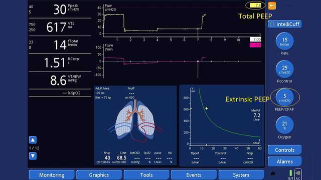
Measuring the total PEEP with an expiratory hold maneuver (see Figure 2 below):

Ensure the Paw waveform is displayed.

  1. Open the Hold window.
  2. Wait until the Paw waveform plot restarts from the left side.
  3. Wait for the next inspiration.
  4. Then select Exp hold. Wait for 3 to 5 seconds, then select Exp hold or press the P&T knob again to deactivate the hold maneuver and close the window.
  5. After the maneuver, the Hold window closes, and the freeze function is activated automatically.
  6. Measure the total PEEP by examining points after flow reached zero on the pressure curve with the cursor.
  7. Calculate AutoPEEP by subtracting extrinsic PEEP from the total PEEP.

Calculations

AutoPEEP = total PEEP - extrinsic PEEP = intrinsic PEEP
PEEP = extrinsic PEEP and is preselected
Total PEEP = intrinsic PEEP + extrinsic PEEP
Screenshot of ventilator display showing expiratory hold maneuver
Figure 2: Measuring total PEEP using an expiratory hold maneuver - Total PEEP of 7.6 cmH2O - extrinsic PEEP of 5 cmH2O = AutoPEEP of 2.6 cmH2O
Screenshot of ventilator display showing expiratory hold maneuver
Figure 2: Measuring total PEEP using an expiratory hold maneuver - Total PEEP of 7.6 cmH2O - extrinsic PEEP of 5 cmH2O = AutoPEEP of 2.6 cmH2O

Avoiding air-trapping

If AutoPEEP is present unintentionally, caregivers should consider adapting the control parameters to avoid air-trapping by increasing the exhalation time. Use of large-diameter endotracheal tubes, bronchodilators, short inspiratory times, long expiratory times, lower respiratory rates and the use of sedatives can be necessary to avoid the dynamic hyperinflation caused by air-trapping.

All Hamilton Medical ventilators feature the intelligent ventilation mode Adaptive Support Ventilation (ASV®). ASV automatically employs lung-protective strategies to minimize complications from AutoPEEP.

Relevant devices: HAMILTON-G5/S1 (sw v2.8x and later); HAMILTON-C3 (sw v2.0.x and later), HAMILTON-C6 (sw v1.1.x and later)

See full citation for (Iotti, G., & Braschi, A. (1999). Measurements of respiratory mechanics during mechanical ventilation. Rhäzüns, Switzerland: Hamilton Medical Scientific Library.1​) below.

Measurements of respiratory mechanics during mechanical ventilation

Iotti, G., & Braschi, A. (1999). Measurements of respiratory mechanics during mechanical ventilation. Rhäzüns, Switzerland: Hamilton Medical Scientific Library.

Invasive mechanical ventilation in COPD and asthma.

García Vicente E, Sandoval Almengor JC, Díaz Caballero LA, Salgado Campo JC. Ventilación mecánica invasiva en EPOC y asma [Invasive mechanical ventilation in COPD and asthma]. Med Intensiva. 2011;35(5):288-298. doi:10.1016/j.medin.2010.11.004

COPD and asthmatic patients use a substantial proportion of mechanical ventilation in the ICU, and their overall mortality with ventilatory support can be significant. From the pathophysiological standpoint, they have increased airway resistance, pulmonary hyperinflation, and high pulmonary dead space, leading to increased work of breathing. If ventilatory demand exceeds work output of the respiratory muscles, acute respiratory failure follows. The main goal of mechanical ventilation in this kind of patients is to improve pulmonary gas exchange and to allow for sufficient rest of compromised respiratory muscles to recover from the fatigued state. The current evidence supports the use of noninvasive positive-pressure ventilation for these patients (especially in COPD), but invasive ventilation also is required frequently in patients who have more severe disease. The physician must be cautious to avoid complications related to mechanical ventilation during ventilatory support. One major cause of the morbidity and mortality arising during mechanical ventilation in these patients is excessive dynamic pulmonary hyperinflation (DH) with intrinsic positive end-expiratory pressure (intrinsic PEEP or auto-PEEP). The purpose of this article is to provide a concise update of the most relevant aspects for the optimal ventilatory management in these patients.

Respiratory mechanics by least squares fitting in mechanically ventilated patients: applications during paralysis and during pressure support ventilation.

Iotti GA, Braschi A, Brunner JX, et al. Respiratory mechanics by least squares fitting in mechanically ventilated patients: applications during paralysis and during pressure support ventilation. Intensive Care Med. 1995;21(5):406-413. doi:10.1007/BF01707409


OBJECTIVE

To evaluate a least squares fitting technique for the purpose of measuring total respiratory compliance (Crs) and resistance (Rrs) in patients submitted to partial ventilatory support, without the need for esophageal pressure measurement.

DESIGN

Prospective, randomized study.

SETTING

A general ICU of a University Hospital.

PATIENTS

11 patients in acute respiratory failure, intubated and assisted by pressure support ventilation (PSV).

INTERVENTIONS

Patients were ventilated at 4 different levels of pressure support. At the end of the study, they were paralyzed for diagnostic reasons and submitted to volume controlled ventilation (CMV).

MEASUREMENTS AND RESULTS

A least squares fitting (LSF) method was applied to measure Crs and Rrs at different levels of pressure support as well as in CMV. Crs and Rrs calculated by the LSF method were compared to reference values which were obtained in PSV by measurement of esophageal pressure, and in CMV by the application of the constant flow, end-inspiratory occlusion method. Inspiratory activity was measured by P0.1. In CMV, Crs and Rrs measured by the LSF method are close to quasistatic compliance (-1.5 +/- 1.5 ml/cmH2O) and to the mean value of minimum and maximum end-inspiratory resistance (+0.9 +/- 2.5 cmH2O/(l/s)). Applied during PSV, the LSF method leads to gross underestimation of Rrs (-10.4 +/- 2.3 cmH2O/(l/s)) and overestimation of Crs (+35.2 +/- 33 ml/cmH2O) whenever the set pressure support level is low and the activity of the respiratory muscles is high (P0.1 was 4.6 +/- 3.1 cmH2O). However, satisfactory estimations of Crs and Rrs by the LSF method were obtained at increased pressure support levels, resulting in a mean error of -0.4 +/- 6 ml/cmH2O and -2.8 +/- 1.5 cmH2O/(l/s), respectively. This condition was coincident with a P0.1 of 1.6 +/- 0.7 cmH2O.

CONCLUSION

The LSF method allows non-invasive evaluation of respiratory mechanics during PSV, provided that a near-relaxation condition is obtained by means of an adequately increased pressure support level. The measurement of P0.1 may be helpful for titrating the pressure support in order to obtain the condition of near-relaxation.