Wir haben festgestellt, dass Sie unsere Website von China aus besuchen.
Für Ihr Land (China) gibt es eine eigene Version der Website.

Zu China wechseln
 Technologien

Transpulmonaler Druck. Die Mechanik des Atemsystems im Klartext

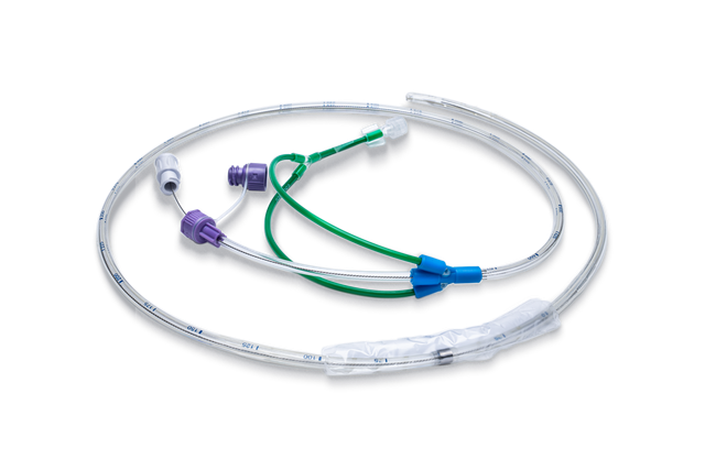
Ösophagealer Druckkatheter NutriVent

Insider-Einblicke. Messung des ösophagealen Drucks

Die Messung des ösophagealen Drucks (Pes) ist eine minimalinvasive Monitoring-Methode, mit der der transpulmonale Druck bestimmt werden kann.

Die gängigste Methode zur Messung des Pes ist die Verwendung eines luftgefüllten Ballons, der in einen Ösophaguskatheter integriert ist.
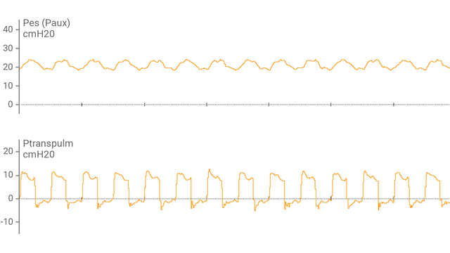
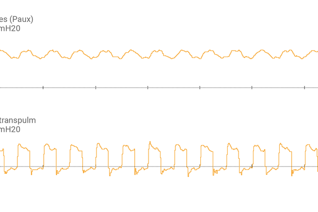
Der Bildschirm des Beatmungsgerätes zeigt den Ösophagusdruck (Pes) und die transpulmonalen Druckwerte (Ptranspulm) als Kurven an. Der Bildschirm des Beatmungsgerätes zeigt den Ösophagusdruck (Pes) und die transpulmonalen Druckwerte (Ptranspulm) als Kurven an.

Mit eigenen Augen sehen. Pes und Ptranspulm auf dem Bildschirm

Nachdem Sie den Ösophagusballonkatheter an den Hilfsanschluss angeschlossen und die korrekte Platzierung bestätigt haben, zeigt der Bildschirm des Beatmungsgerätes den Ösophagusdruck (Pes) und die transpulmonalen Druckwerte (Ptranspulm) als Kurven an.

Sie können den statischen Ptranspulm mit inspiratorischen und exspiratorischen Hold-Manövern messen.
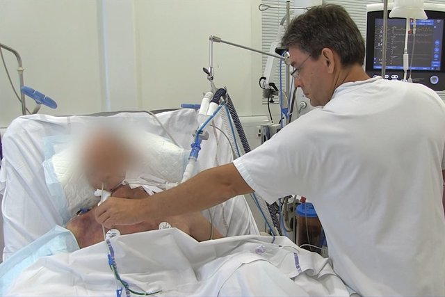
Arzt mit einem Patienten

Gemeinsam an einem Strang. Beurteilung der Lungenrekrutierbarkeit

Zur Beurteilung der Rekrutierbarkeit und zur Durchführung von Recruitmentmanövern kann der transpulmonale Druck auch in Kombination mit dem P/V Tool® verwendet werden.
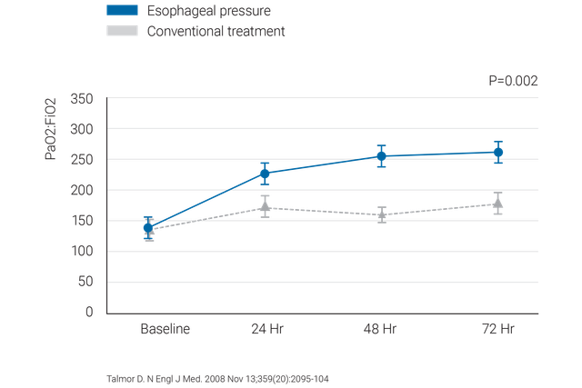
Statistische Grafik: Talmor D. N Engl J Med. 2008 Nov 13;359(20):2095-104

Was spricht dafür? Klinische Nachweise im Überblick

  • Die Einstellung von PEEP anhand des transpulmonalen Drucks verbesserte die Compliance und Oxygenierung bei ARDS-Patienten (Talmor D, Sarge T, Malhotra A, et al. Mechanical ventilation guided by esophageal pressure in acute lung injury. N Engl J Med. 2008;359(20):2095-2104. doi:10.1056/NEJMoa07086381​).
  • Eine gezielte Messung des positiven transpulmonaren Drucks verbesserte die Elastance und den Driving Pressure und kann mit einer verbesserten 28-Tage-Mortalität in Verbindung gebracht werden (Baedorf Kassis E, Loring SH, Talmor D. Mortality and pulmonary mechanics in relation to respiratory system and transpulmonary driving pressures in ARDS. Intensive Care Med. 2016;42(8):1206-1213. doi:10.1007/s00134-016-4403-72​).
  • Durch das transpulmonale Druck-Monitoring kann auch bei schwerstkranken Patienten der Einsatz eines extrakorporalen Membranoxygenierungsgerätes (ECMO) vermieden werden (Grasso S, Terragni P, Birocco A, et al. ECMO criteria for influenza A (H1N1)-associated ARDS: role of transpulmonary pressure. Intensive Care Med. 2012;38(3):395-403. doi:10.1007/s00134-012-2490-73​).
  • Eine Beatmungsstrategie, die sich am transpulmonalen Druck orientiert, kann den Anteil der Patienten mit schwerem ARDS erhöhen, die erfolgreich von der ECMO entwöhnt werden (Wang R, Sun B, Li X, et al. Mechanical Ventilation Strategy Guided by Transpulmonary Pressure in Severe Acute Respiratory Distress Syndrome Treated With Venovenous Extracorporeal Membrane Oxygenation. Crit Care Med. 2020;48(9):1280-1288. doi:10.1097/CCM.00000000000044454​).
  • Die Technik der Pes-Messung ist der Goldstandard für die Bewertung der Atemanstrengung und der Atemarbeit (Bertoni M, Spadaro S, Goligher EC. Monitoring Patient Respiratory Effort During Mechanical Ventilation: Lung and Diaphragm-Protective Ventilation. Crit Care. 2020;24(1):106. Published 2020 Mar 24. doi:10.1186/s13054-020-2777-y5​).
Grafische Darstellung: Student hält sein Diplom in der Hand

Gut zu wissen! Schulungsressourcen für den transpulmonalen Druck

Watch this short demonstration to learn how to use transpulmonary pressure measurement to set PEEP in mechanically ventilated patients more accurately.

Einstellen von PEEP mit dem transpulmonalen Druck

Sehen Sie sich diese kurze Demonstration an, um zu verstehen, wie Sie mit dem transpulmonalen Druck-Monitoring den PEEP genauer einstellen können.

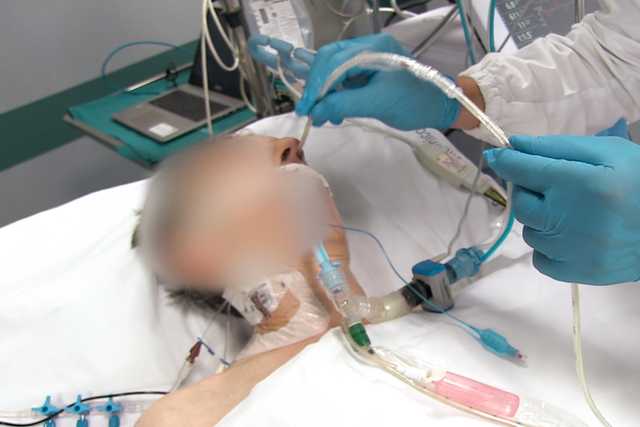
Einführen eines ösophagealen Ballonkatheters in den Patienten

Es ist alles eine Frage der Technik! Einführen eines ösophagealen Ballonkatheters

13 Expertentipps (eBook)

Kostenloses eBook

13 Expertentipps. Messung des ösophagealen Drucks

Bewährte Empfehlungen aus der klinischen Praxis, wie Sie bei der Verwendung des ösophagealen Drucks bei ARDS-Patienten vorgehen und was Sie vermeiden sollten.

Verbrauchsmaterialien

Wir bieten ösophageale Ballonkatheter von CooperSurgical und nasogastrale NutriVent-Katheter.

Verfügbarkeit

Das transpulmonale Druck-Monitoring zählt zur Standardausstattung bei den Beatmungsgeräten HAMILTON-C6 und HAMILTON-G5/S1.

Mechanical ventilation guided by esophageal pressure in acute lung injury.

Talmor D, Sarge T, Malhotra A, et al. Mechanical ventilation guided by esophageal pressure in acute lung injury. N Engl J Med. 2008;359(20):2095-2104. doi:10.1056/NEJMoa0708638


BACKGROUND

Survival of patients with acute lung injury or the acute respiratory distress syndrome (ARDS) has been improved by ventilation with small tidal volumes and the use of positive end-expiratory pressure (PEEP); however, the optimal level of PEEP has been difficult to determine. In this pilot study, we estimated transpulmonary pressure with the use of esophageal balloon catheters. We reasoned that the use of pleural-pressure measurements, despite the technical limitations to the accuracy of such measurements, would enable us to find a PEEP value that could maintain oxygenation while preventing lung injury due to repeated alveolar collapse or overdistention.

METHODS

We randomly assigned patients with acute lung injury or ARDS to undergo mechanical ventilation with PEEP adjusted according to measurements of esophageal pressure (the esophageal-pressure-guided group) or according to the Acute Respiratory Distress Syndrome Network standard-of-care recommendations (the control group). The primary end point was improvement in oxygenation. The secondary end points included respiratory-system compliance and patient outcomes.

RESULTS

The study reached its stopping criterion and was terminated after 61 patients had been enrolled. The ratio of the partial pressure of arterial oxygen to the fraction of inspired oxygen at 72 hours was 88 mm Hg higher in the esophageal-pressure-guided group than in the control group (95% confidence interval, 78.1 to 98.3; P=0.002). This effect was persistent over the entire follow-up time (at 24, 48, and 72 hours; P=0.001 by repeated-measures analysis of variance). Respiratory-system compliance was also significantly better at 24, 48, and 72 hours in the esophageal-pressure-guided group (P=0.01 by repeated-measures analysis of variance).

CONCLUSIONS

As compared with the current standard of care, a ventilator strategy using esophageal pressures to estimate the transpulmonary pressure significantly improves oxygenation and compliance. Multicenter clinical trials are needed to determine whether this approach should be widely adopted. (ClinicalTrials.gov number, NCT00127491.)

Mortality and pulmonary mechanics in relation to respiratory system and transpulmonary driving pressures in ARDS.

Baedorf Kassis E, Loring SH, Talmor D. Mortality and pulmonary mechanics in relation to respiratory system and transpulmonary driving pressures in ARDS. Intensive Care Med. 2016;42(8):1206-1213. doi:10.1007/s00134-016-4403-7


PURPOSE

The driving pressure of the respiratory system has been shown to strongly correlate with mortality in a recent large retrospective ARDSnet study. Respiratory system driving pressure [plateau pressure-positive end-expiratory pressure (PEEP)] does not account for variable chest wall compliance. Esophageal manometry can be utilized to determine transpulmonary driving pressure. We have examined the relationships between respiratory system and transpulmonary driving pressure, pulmonary mechanics and 28-day mortality.

METHODS

Fifty-six patients from a previous study were analyzed to compare PEEP titration to maintain positive transpulmonary end-expiratory pressure to a control protocol. Respiratory system and transpulmonary driving pressures and pulmonary mechanics were examined at baseline, 5 min and 24 h. Analysis of variance and linear regression were used to compare 28 day survivors versus non-survivors and the intervention group versus the control group, respectively.

RESULTS

At baseline and 5 min there was no difference in respiratory system or transpulmonary driving pressure. By 24 h, survivors had lower respiratory system and transpulmonary driving pressures. Similarly, by 24 h the intervention group had lower transpulmonary driving pressure. This decrease was explained by improved elastance and increased PEEP.

CONCLUSIONS

The results suggest that utilizing PEEP titration to target positive transpulmonary pressure via esophageal manometry causes both improved elastance and driving pressures. Treatment strategies leading to decreased respiratory system and transpulmonary driving pressure at 24 h may be associated with improved 28 day mortality. Studies to clarify the role of respiratory system and transpulmonary driving pressures as a prognosticator and bedside ventilator target are warranted.

ECMO criteria for influenza A (H1N1)-associated ARDS: role of transpulmonary pressure.

Grasso S, Terragni P, Birocco A, et al. ECMO criteria for influenza A (H1N1)-associated ARDS: role of transpulmonary pressure. Intensive Care Med. 2012;38(3):395-403. doi:10.1007/s00134-012-2490-7


PURPOSE

To assess whether partitioning the elastance of the respiratory system (E (RS)) between lung (E (L)) and chest wall (E (CW)) elastance in order to target values of end-inspiratory transpulmonary pressure (PPLAT(L)) close to its upper physiological limit (25 cmH(2)O) may optimize oxygenation allowing conventional treatment in patients with influenza A (H1N1)-associated ARDS referred for extracorporeal membrane oxygenation (ECMO).

METHODS

Prospective data collection of patients with influenza A (H1N1)-associated ARDS referred for ECMO (October 2009-January 2010). Esophageal pressure was used to (a) partition respiratory mechanics between lung and chest wall, (b) titrate positive end-expiratory pressure (PEEP) to target the upper physiological limit of PPLAT(L) (25 cmH(2)O).

RESULTS

Fourteen patients were referred for ECMO. In seven patients PPLAT(L) was 27.2 ± 1.2 cmH(2)O; all these patients underwent ECMO. In the other seven patients, PPLAT(L) was 16.6 ± 2.9 cmH(2)O. Raising PEEP (from 17.9 ± 1.2 to 22.3 ± 1.4 cmH(2)O, P = 0.0001) to approach the upper physiological limit of transpulmonary pressure (PPLAT(L) = 25.3 ± 1.7 cm H(2)O) improved oxygenation index (from 37.4 ± 3.7 to 16.5 ± 1.4, P = 0.0001) allowing patients to be treated with conventional ventilation.

CONCLUSIONS

Abnormalities of chest wall mechanics may be present in some patients with influenza A (H1N1)-associated ARDS. These abnormalities may not be inferred from measurements of end-inspiratory plateau pressure of the respiratory system (PPLAT(RS)). In these patients, titrating PEEP to PPLAT(RS) may overestimate the incidence of hypoxemia refractory to conventional ventilation leading to inappropriate use of ECMO.

Mechanical Ventilation Strategy Guided by Transpulmonary Pressure in Severe Acute Respiratory Distress Syndrome Treated With Venovenous Extracorporeal Membrane Oxygenation.

Wang R, Sun B, Li X, et al. Mechanical Ventilation Strategy Guided by Transpulmonary Pressure in Severe Acute Respiratory Distress Syndrome Treated With Venovenous Extracorporeal Membrane Oxygenation. Crit Care Med. 2020;48(9):1280-1288. doi:10.1097/CCM.0000000000004445


OBJECTIVES

Previous studies have suggested that adjusting ventilator settings based on transpulmonary pressure measurements may minimize ventilator-induced lung injury, but this has never been investigated in patients with severe acute respiratory distress syndrome supported with venovenous extracorporeal membrane oxygenation. We aimed to evaluate whether a transpulmonary pressure-guided ventilation strategy would increase the proportion of patients successfully weaned from venovenous extracorporeal membrane oxygenation support in patients with severe acute respiratory distress syndrome.

DESIGN

Single-center, prospective, randomized controlled trial.

SETTING

Sixteen-bed, respiratory ICU at a tertiary academic medical center.

PATIENTS

Severe acute respiratory distress syndrome patients receiving venovenous extracorporeal membrane oxygenation.

INTERVENTIONS

One-hundred four patients were randomized to transpulmonary pressure-guided ventilation group (n = 52) or lung rest strategy group (n = 52) groups. Two patients had cardiac arrest during establishment of venovenous extracorporeal membrane oxygenation in the lung rest group did not receive the assigned intervention. Thus, 102 patients were included in the analysis.

MEASUREMENTS AND MAIN RESULTS

The proportion of patients successfully weaned from venovenous extracorporeal membrane oxygenation in the transpulmonary pressure-guided group was significantly higher than that in the lung rest group (71.2% vs 48.0%; p = 0.017). Compared with the lung rest group, driving pressure, tidal volumes, and mechanical power were significantly lower, and positive end-expiratory pressure was significantly higher, in the transpulmonary pressure-guided group during venovenous extracorporeal membrane oxygenation support. In the transpulmonary pressure-guided group, levels of interleukin-1β, interleukin-6, and interleukin-8 were significantly lower, and interleukin-10 was significantly higher, than those of the lung rest group over time. Lung density was significantly lower in the transpulmonary pressure-guided group after venovenous extracorporeal membrane oxygenation support than in the lung rest group.

CONCLUSIONS

A transpulmonary pressure-guided ventilation strategy could increase the proportion of patients with severe acute respiratory distress syndrome successfully weaned from venovenous extracorporeal membrane oxygenation.

Monitoring Patient Respiratory Effort During Mechanical Ventilation: Lung and Diaphragm-Protective Ventilation.

Bertoni M, Spadaro S, Goligher EC. Monitoring Patient Respiratory Effort During Mechanical Ventilation: Lung and Diaphragm-Protective Ventilation. Crit Care. 2020;24(1):106. Published 2020 Mar 24. doi:10.1186/s13054-020-2777-y

This article is one of ten reviews selected from the Annual Update in Intensive Care and Emergency Medicine 2020. Other selected articles can be found online at https://www.biomedcentral.com/collections/annualupdate2020. Further information about the Annual Update in Intensive Care and Emergency Medicine is available from http://www.springer.com/series/8901.