Wir haben festgestellt, dass Sie unsere Website von China aus besuchen.
Für Ihr Land (China) gibt es eine eigene Version der Website.

Zu China wechseln
 Verbrauchsmaterialien

Flow-Sensoren. Proximale Flow- und Druckmessung

Flow-Sensor

Kommen Sie näher! Proximale Flow-Messung

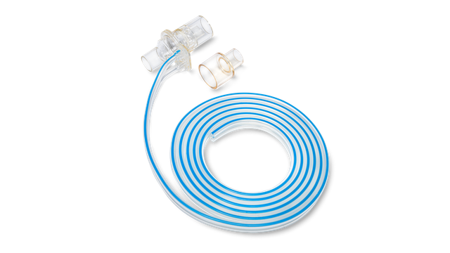
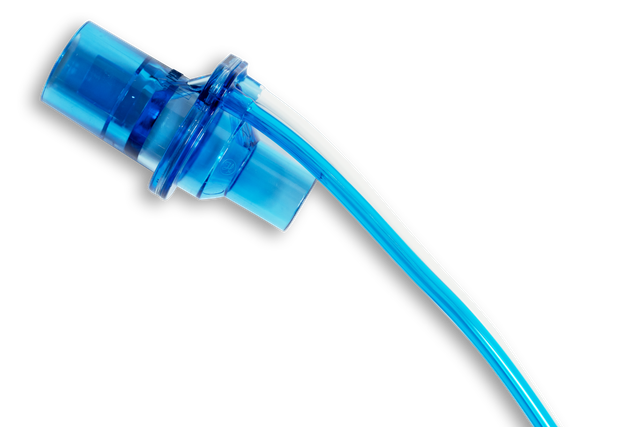
Der proximale Flow-Sensor ist seit 1983 das Herzstück unserer Beatmungsgeräte. Der gesamte Beatmungsprozess hängt von der Messung und Genauigkeit des Flow-Sensors ab, der auch Daten von der Atemwegsöffnung liefert.

Präzise Volumen-, Flow- und Druckdaten sind entscheidend für eine korrekte Diagnose und die Vermeidung von häufigen Nebenwirkungen aufgrund ungeeigneter Beatmungseinstellungen. Sie ermöglichen auch die Anwendung einiger unserer hochmodernen Technologien, wie z. B. die Modi ASV und INTELLiVENT-ASV, IntelliSync+ und das P/V-Tool.

Flow-Sensor

Genauigkeit ist ein Muss. Das Wohl Ihrer Patienten steht auf dem Spiel

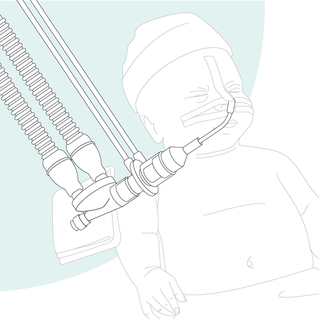
Unsere Beatmungsgeräte messen Flow und Druck nahe am Atemweg des Patienten. Studien haben gezeigt, dass die Tidalvolumina für beatmete Patienten mit einem am Endotrachealtubus platzierten Flow-Sensor bestimmt werden sollten (Cannon ML, Cornell J, Tripp-Hamel DS, et al. Tidal volumes for ventilated infants should be determined with a pneumotachometer placed at the endotracheal tube. Am J Respir Crit Care Med. 2000;162(6):2109-2112. doi:10.1164/ajrccm.162.6.99061121​, Gammage, Gary W.; Banner, Michael J.; Blanch, Paul B.; Kirby, Robert R. VENTILATOR DISPLAYED TIDAL VOLUME—WHAT YOU SEE MAY NOT BE WHAT YOU GET, Critical Care Medicine: April 1988 - Volume 16 - Issue 4 - p 454 2​).

Grafische Darstellung: eine Frau, die über eine Frage nachdenkt

Gibt es Beweise? Klinische Nachweise

Die genaue Bestimmung des ausgeatmeten Tidalvolumens (VTE) ist von entscheidender Bedeutung (Cannon ML, Cornell J, Tripp-Hamel DS, et al. Tidal volumes for ventilated infants should be determined with a pneumotachometer placed at the endotracheal tube. Am J Respir Crit Care Med. 2000;162(6):2109-2112. doi:10.1164/ajrccm.162.6.99061121​), insbesondere in Situationen, in denen nur kleine Tidalvolumina abgegeben werden (Kleinkinder, Neonaten und ARDS-Patienten). Mit den Flow-Sensoren von Hamilton Medical können Sie das VTE in der Nähe der Atemwege des Patienten messen, um einen präziseren Wert zu erhalten.

Ihre Vorteile:

  • Durch die proximale Platzierung werden die Auswirkungen der Beatmungsschlauchsystem-Compliance auf die Flow- und Volumenmessungen eliminiert (Cannon ML, Cornell J, Tripp-Hamel DS, et al. Tidal volumes for ventilated infants should be determined with a pneumotachometer placed at the endotracheal tube. Am J Respir Crit Care Med. 2000;162(6):2109-2112. doi:10.1164/ajrccm.162.6.99061121​)
  • Bei der VTE-Messung ist die Resistance des Atemsystems geringer (Nève V, Leclerc F, Noizet O, et al. Influence of respiratory system impedance on volume and pressure delivered at the Y piece in ventilated infants. Pediatr Crit Care Med. 2003;4(4):418-425. doi:10.1097/01.PCC.0000090289.98377.153​)
  • Es treten weniger Leckagen auf, die das Ergebnis verfälschen könnten (Al-Majed SI, Thompson JE, Watson KF, Randolph AG. Effect of lung compliance and endotracheal tube leakage on measurement of tidal volume. Crit Care. 2004;8(6):R398-R402. doi:10.1186/cc29544​)

Unser Portfolio an Flow-Sensoren

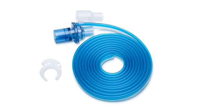
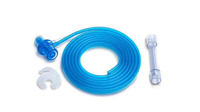
Wir bieten Verbrauchsmaterialien von Hamilton Medical für erwachsene, pädiatrische und neonatale Patienten. Je nach den Richtlinien in Ihrer Einrichtung haben Sie die Wahl zwischen wiederverwendbaren Produkten und Ausführungen für den Einmalgebrauch.
Dr. Robert Lopez

Kundenstimmen

Mit den Flow-Sensoren für den Einmalgebrauch von Hamilton Medical müssen wir uns keine Gedanken mehr zu einer Kreuzkontamination machen, da wir denselben Flow-Sensor nicht bei einem anderen Patienten einsetzen.

Dr. Robert Lopez

Leiter der Beatmungsstation bis 2018
University Medical Center, Lubbock (TX), USA
Tidal volumes for ventilated infants should be determined with a pneumotachometer placed at the endotracheal tube.

Cannon ML, Cornell J, Tripp-Hamel DS, et al. Tidal volumes for ventilated infants should be determined with a pneumotachometer placed at the endotracheal tube. Am J Respir Crit Care Med. 2000;162(6):2109-2112. doi:10.1164/ajrccm.162.6.9906112

Many ventilators measure expired tidal volume (VT) without compensation either for the compliance of the ventilator circuit or for variations in the circuit setup. We hypothesized that the exhaled VT measured with a conventional ventilator at the expiratory valve would differ significantly from the exhaled VT measured with a pneumotachometer placed at the endotracheal tube. To investigate this we studied 98 infants and children requiring conventional ventilation. We used linear regression analysis to compare the VT obtained with the pneumotachometer with the ventilator-measured volume. An additional comparison was made between the pneumotachometer volume and a calculated effective VT. For infant circuits (n = 70), our analysis revealed a poor correlation between the expiratory VT measured with the pneumotachometer and the ventilator-measured volume (r(2) = 0.54). Similarly, the expiratory VT measured with the pneumotachometer did not correlate with the calculated effective volume (r(2) = 0.58). For pediatric circuits (n = 28), there was improved correlation between the expiratory VT measured with the pneumotachometer and both the ventilator-measured volume and the calculated effective VT (r(2) = 0.84 and r(2) = 0.85, respectively). The data demonstrate a significant discrepancy between expiratory VT measured at a ventilator and that measured with a pneumotachometer placed at the endotracheal tube in infants. Correcting for the compliance of the ventilator circuit by calculating the effective VT did not alter this discrepancy. In conventionally ventilated infants, exhaled VT should be determined with a pneumotachometer placed at the airway.

Ventilator displayed tidal volume: What you see may not be what you get.

Gammage, Gary W.; Banner, Michael J.; Blanch, Paul B.; Kirby, Robert R. VENTILATOR DISPLAYED TIDAL VOLUME—WHAT YOU SEE MAY NOT BE WHAT YOU GET, Critical Care Medicine: April 1988 - Volume 16 - Issue 4 - p 454

Influence of respiratory system impedance on volume and pressure delivered at the Y piece in ventilated infants.

Nève V, Leclerc F, Noizet O, et al. Influence of respiratory system impedance on volume and pressure delivered at the Y piece in ventilated infants. Pediatr Crit Care Med. 2003;4(4):418-425. doi:10.1097/01.PCC.0000090289.98377.15


OBJECTIVES

Tidal volume (VT) delivered to infants' airways are overestimated and pressure underestimated when measured in the ventilator and not at the Y piece. This study aimed at evaluating the influence of respiratory system impedance on expiratory VT (VTE) and pressure measurement difference.

DESIGN

Prospective observational study.

SETTING

Pediatric intensive care unit at a university hospital.

PATIENTS

Data were collected between February 2000 and October 2001 for 30 infants (range, 1-23 months) ventilated in the pressure-controlled or volume-controlled mode.

INTERVENTIONS

Measurements of VTE, pressure obtained at the same time at the Y piece and on the ventilator Servo 300, were collected in ventilated infants. Respiratory system impedance was calculated from data obtained at the Y piece. Circuit compliance was measured in vitro. VTEs were corrected for compressible volume.

MEASUREMENTS AND RESULTS

VTEs were overestimated by the Servo 300 in the pressure-controlled and volume-controlled modes (from 5% to 62% of the value displayed on Servo 300). Maximal inspiratory pressures were underestimated by the Servo 300 in the pressure-controlled mode (difference from -2 to +19 cm H(2)O). Measurement difference increased with increasing respiratory system impedance. Ventilator VTE corrected for circuit compliance did not offer a sufficiently accurate estimation of VTE at the Y piece.

CONCLUSIONS

VT and pressure measurements must be performed at the Y piece, especially in infants with increased respiratory system impedance (i.e., decreased respiratory system compliance or increased resistance). Correcting VTE for circuit compliance cannot replace measurement of VT at the Y piece.

Effect of lung compliance and endotracheal tube leakage on measurement of tidal volume.

Al-Majed SI, Thompson JE, Watson KF, Randolph AG. Effect of lung compliance and endotracheal tube leakage on measurement of tidal volume. Crit Care. 2004;8(6):R398-R402. doi:10.1186/cc2954


INTRODUCTION

The objective of this laboratory study was to measure the effect of decreased lung compliance and endotracheal tube (ETT) leakage on measured exhaled tidal volume at the airway and at the ventilator, in a research study with a test lung.

METHODS

The subjects were infant, adult and pediatric test lungs. In the test lung model, lung compliances were set to normal and to levels seen in acute respiratory distress syndrome. Set tidal volume was 6 ml/kg across a range of simulated weights and ETT sizes. Data were recorded from both the ventilator light-emitting diode display and the CO2SMO Plus monitor display by a single observer. Effective tidal volume was calculated from a standard equation.

RESULTS

In all test lung models, exhaled tidal volume measured at the airway decreased markedly with decreasing lung compliance, but measurement at the ventilator showed minimal change. In the absence of a simulated ETT leak, calculation of the effective tidal volume led to measurements very similar to exhaled tidal volume measured at the ETT. With a simulated ETT tube leak, the effective tidal volume markedly overestimated tidal volume measured at the airway.

CONCLUSION

Previous investigators have emphasized the need to measure tidal volume at the ETT for all children. When ETT leakage is minimal, it seems from our simulated lung models that calculation of effective tidal volume would give similar readings to tidal volume measured at the airway, even in small patients. Future studies of tidal volume measurement accuracy in mechanically ventilated children should control for the degree of ETT leakage.