Zurück

How to manage cuff pressure in children intubated with cuffed tubes

Artikel

Autor: Süha Demirakca

Datum: 07.11.2019

The choice of tube size for uncuffed tubes is commonly made according to Cole`s formula (Age/4 + 4 mm); for cuffed tubes a size 0.5 mm smaller is recommended. The cuff pressure needs to be sufficient to avoid leakage during mechanical ventilation, and also to completely seal the airway and prevent micro-aspiration (thus preventing ventilator-associated pneumonia).
How to manage cuff pressure in children intubated with cuffed tubes

Minimal leakage at peak inspiratory pressure

However over-inflation, particularly over a prolonged period, may limit mucosal perfusion. The standard cuff pressure to provide leakage-free ventilation is usually set at 25 mbar. Absence of leakage is primarily essential in special situations such as during a recruitment maneuver. In all other circumstances, common practice in many PICU`s is to keep the cuff inflated to provide minimal leakage at peak inspiratory pressure, so that cuff pressure can usually be down-titrated to values between 20–25 mbar.

Leak thresholds

Cuff pressure leak thresholds in children will vary with sedation status and positioning of the patient, and are also influenced by coughing and suctioning. Pressure drops occur within a few hours after manual adjustment of the cuff pressure. Therefore, regular monitoring of the cuff pressure to provide accurate cuff pressure control is very important. This can be best achieved with an online cuff controller tool such as Intellicuff.

A pre-extubation leak pressure test may provide information about the risk of a subglottic upper airway obstruction occurring. Because the cuff is located in the subglottic area, the cuff leak pressure threshold appears to measure subglottic edema. A simple method of gaining this information is to test whether an audible leak occurs at a peak inspiratory pressure of 25 mbar, after the patient has been suctioned and the cuff has been deflated. The presence of an audible pre-extubation leak at 25 mbar indicates that no subglottic upper airway obstruction will occur, whereas the absence of leak at 25 mbar signals a subglottic stenosis that will need treatment in 18%–19% of patients. The importance of leakage in the case of cuffed endotracheal tubes may be related to the fact that in comparison to uncuffed tubes, the cuffed tubes used are usually smaller because the cuff can be inflated to maintain tidal volume and pressure.

Full citataions see below: Herbinger LA. Evidence Based Use of Cuffed Endotracheal Tubes in Children. J Perianesth Nurs. 2018;33(5):590-600. doi:10.1016/j.jopan.2017.07.0011​, Calder A, Hegarty M, Erb TO, von Ungern-Sternberg BS. Predictors of postoperative sore throat in intubated children. Paediatr Anaesth. 2012;22(3):239-243. doi:10.1111/j.1460-9592.2011.03727.x2​, Newth CJ, Rachman B, Patel N, Hammer J. The use of cuffed versus uncuffed endotracheal tubes in pediatric intensive care. J Pediatr. 2004;144(3):333-337. doi:10.1016/j.jpeds.2003.12.0183​, Schneider J, Mulale U, Yamout S, Pollard S, Silver P. Impact of monitoring endotracheal tube cuff leak pressure on postextubation stridor in children. J Crit Care. 2016;36:173-177. doi:10.1016/j.jcrc.2016.06.0334​, Babic SA and Chatburn RL. Laboratory Evaluation of Cuff Pressure Control Methods. Respir Care. 2019 Jul 30. pii: respcare.06728. doi: 10.4187/respcare.06728. [Epub ahead of print]5​, Khemani RG, Hotz J, Morzov R, et al. Evaluating Risk Factors for Pediatric Post-extubation Upper Airway Obstruction Using a Physiology-based Tool. Am J Respir Crit Care Med. 2016;193(2):198-209. doi:10.1164/rccm.201506-1064OC6​, Chung YH, Chao TY, Chiu CT, Lin MC. The cuff-leak test is a simple tool to verify severe laryngeal edema in patients undergoing long-term mechanical ventilation. Crit Care Med. 2006;34(2):409-414. doi:10.1097/01.ccm.0000198105.65413.857
Screenshot showing IntelliCuff set to auto and cuff pressure limits
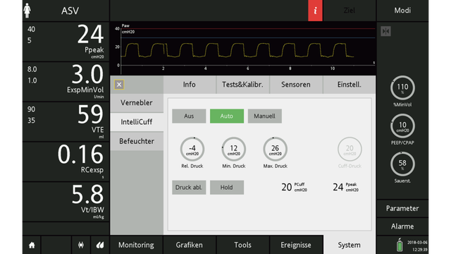
Figure 1: An 18-month old child ventilated with a Ppeak of 24 cmH2O (PEEP 10 cmH2O). IntelliCuff is set to auto control. The cuff pressure is set at -4 cmH2O in relation to Ppeak. The cuff pressure lower limit is 12 cmH2O; the upper limit 26 cmH2O.
Screenshot showing IntelliCuff set to manual
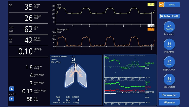
Figure 2: An 8-year old child with severe ARDS ventilated with a Ppeak of 35 cmH2O and PEEP of 22 cmH2O. IntelliCuff is set to manual control. The cuff pressure is titrated to 23 cmH2O to allow a minimum leak (4%).
Screenshot showing IntelliCuff set to manual
Figure 2: An 8-year old child with severe ARDS ventilated with a Ppeak of 35 cmH2O and PEEP of 22 cmH2O. IntelliCuff is set to manual control. The cuff pressure is titrated to 23 cmH2O to allow a minimum leak (4%).

Evidence Based Use of Cuffed Endotracheal Tubes in Children.

Herbinger LA. Evidence Based Use of Cuffed Endotracheal Tubes in Children. J Perianesth Nurs. 2018;33(5):590-600. doi:10.1016/j.jopan.2017.07.001

Historically, the use of cuffed endotracheal tubes (ETTs) was reserved for children aged 8 years or older to minimize the risks of postextubation laryngeal edema. However, since publication of a 1997 study, researchers have consistently presented evidence that appropriately used cuffed ETTs are as safe as uncuffed ETTs. Because of the advantages of cuffed ETTs in the perianesthesia setting, the transition to cuffed ETTs in children is now complete. However, risks related to using cuffed ETTs in young children increase when guidelines for safe and appropriate use are not followed. Perianesthesia practitioners caring for children must understand the implications related to ETT type, correct ETT sizing, and the monitoring and control of ETT cuff pressure. The purpose of this educational module is to present evidence-based guidelines for the appropriate use of cuffed ETTs in children less than 8 years of age in the perianesthesia setting.

Predictors of postoperative sore throat in intubated children.

Calder A, Hegarty M, Erb TO, von Ungern-Sternberg BS. Predictors of postoperative sore throat in intubated children. Paediatr Anaesth. 2012;22(3):239-243. doi:10.1111/j.1460-9592.2011.03727.x


BACKGROUND

The incidence of postoperative sore throat (POST) following intubation is not well defined in the pediatric population. The etiology is multifactorial and includes impairment of subglottic mucosal perfusion and edema as a result of the pressures exerted by cuffed or uncuffed tubes.

AIM

To determine the incidence of, and risk factors for, POST in intubated children undergoing elective day-case surgery.

METHODS

Five hundred patients aged 3-16 years were studied prospectively. Endotracheal tube (ETT) choice (cuffed or uncuffed) was left to the anesthetist. The cuff was inflated either until loss of audible leak or to a determined pressure using a cuff manometer. The research team then measured the cuff pressure (CP). POST incidence and intensity was determined by interviewing patients prior to discharge from the same day procedure unit. Chi-square testing and stepwise logistic regression were used to determine the predictors of POST.

RESULTS

Of the 111 (22%) children developed a sore throat, 19 (3.8%) a sore neck, and 5 (1%) a sore jaw. 19% of patients with cuffed ETTs complained of sore throat compared with 37% of those intubated with an uncuffed ETT. The incidence of POST increased with CP; 0-10% at 0 cmH(2)O, 4% at 11-20 cmH(2)O, 20% at 21-30 cmH(2)O, 68% at CP 31-40 cmH(2)O, and 96% at CP >40 cmH(2)O. The ETT CP and use of uncuffed ETTs were univariate predictors of POST.

CONCLUSIONS

Children intubated with uncuffed ETTs are more likely to have POST. ETT CP is positively correlated with the incidence of POST. When using cuffed ETTs, CP should be routinely measured intraoperatively.

The use of cuffed versus uncuffed endotracheal tubes in pediatric intensive care.

Newth CJ, Rachman B, Patel N, Hammer J. The use of cuffed versus uncuffed endotracheal tubes in pediatric intensive care. J Pediatr. 2004;144(3):333-337. doi:10.1016/j.jpeds.2003.12.018


OBJECTIVE

To report our experience with cuffed endotracheal tubes (ETT) in a large cohort of critically ill children. Study design We prospectively collected data over a 1-year period concerning long-term intubation on 860 critically ill children admitted to our intensive care unit. Tube sizes were dictated by the modified Cole formula for uncuffed ETT (age [y]/4+4 mm ID) and chosen one-half size less for cuffed ETT. Cuff pressure was regularly monitored to maintain a small leak at peak inspiratory pressure. The choice of ETT was made by the physician responsible for the initial airway management.

RESULTS

There were 597 patients in the first 5 years of life, with 210 having cuffed ETT. There were no significant differences in the use of racemic epinephrine for postextubation subglottic edema, the rate of successful extubation or the need for tracheotomy between those with cuffed and uncuffed ETT in any age group.

CONCLUSIONS

Our data suggest that the traditional teaching in pediatric anesthesia and intensive care, including current pediatric life support recommendations, need to be reviewed for children to benefit from the advantages of modern low-pressure cuffed ETT during critical illnesses.

Impact of monitoring endotracheal tube cuff leak pressure on postextubation stridor in children.

Schneider J, Mulale U, Yamout S, Pollard S, Silver P. Impact of monitoring endotracheal tube cuff leak pressure on postextubation stridor in children. J Crit Care. 2016;36:173-177. doi:10.1016/j.jcrc.2016.06.033


PURPOSE

To determine if implementing a protocol maintaining an air leak when using cuffed endotracheal tubes (ETT) throughout the course of mechanical ventilation (MV) in children would decrease the rate of postextubation stridor (PES).

METHODS

All children requiring MV through a cuffed ETT were included, except those with (1) upper airway anomaly, (2) died while on MV, (3) received tracheostomy before extubation, and (4) transferred before extubation. We implemented a protocol limiting the volume of air instilled into the cuff, allowing an air leak by 25 cm H2O pressure or by peak inspiratory pressure, whichever was higher. Monitoring occurred every 6 hours, adjusting cuff volumes if necessary. Patients receiving nebulized racemic epinephrine within 24 hours of extubation for upper airway obstruction were defined as having PES.

RESULTS

At baseline, 110 patients received cuffed ETTs. The proportion of patients who had an air leak at the time of extubation was 47.3%, and that who developed PES was 21.8%. During the intervention, 101 patients received cuffed ETTs. Most (72.3%) had an air leak at the time of extubation (P< .01), and 9.9% developed PES, a 54.6% relative decrease (relative risk, 0.45; 95% confidence interval, 0.22-0.90; P= .018).

CONCLUSIONS

Maintaining an appropriate air leak throughout the course of MV using cuffed ETT decreases the rate of PES in children.

Laboratory Evaluation of Cuff Pressure Control Methods.

Babic SA, Chatburn RL. Laboratory Evaluation of Cuff Pressure Control Methods. Respir Care. 2020;65(1):62-67. doi:10.4187/respcare.06728


BACKGROUND

Automatic cuff pressure (Pcuff) control devices for artificial airways are available, yet there are no standards or data to support their use. We hypothesized that airway pressure oscillations during mechanical ventilation are transmitted to Pcuff; and that the change in mean Pcuff (ΔPcuff) is zero during mechanical ventilation with controlled or uncontrolled Pcuff.

METHODS

Experiments lasted 12 h, and 2 inspiratory pressure targets (Pinsp) were established. We tested 3 automatic devices (Intellicuff Standalone, PressureEyes, and Tracoe) and one manual method for uncontrolled Pcuff. We utilized a training mannequin with an 8-mm endotracheal tube to assess pressure-controlled continuous mechanical ventilation with the following parameters: breathing frequency = 20 breaths/min, TI = 1.0 s, PEEP = 10 cm H2O, and Pinsp = 10 and 40 cm H2O. For automatic cuff pressure control, we used a data acquisition system. For manual cuff pressure control, Pcuff was set once and measured after mechanical ventilation. Initial Pcuff was 25 cm H2O, and ΔPcuff was calculated as final mean Pcuff - initial mean Pcuff. Data for ΔPcuff were compared with t tests and reported as mean (SD).

RESULTS

Airway pressure oscillations during ventilation were observed in Pcuff waveforms. For manual control, ΔPcuff was -9.3 (2.1) cm H2O for Pinsp = 10 cm H2O and -8.1 (1.1) cm H2O for Pinsp = 40 cm H2O (vs 0, P < .001). There was no difference in ΔPcuff for Pinsp = 10 cm H2O versus 40 cm H2O (P = .21). ΔPcuff was only ± 0.3 cm H2O for automatic control, which we deemed clinically unimportant.

CONCLUSIONS

Automatic devices do not regulate ventilatory pressure oscillations, but they do control mean Pcuff and keep ΔPcuff well below a clinically important threshold. The large ΔPcuff seen with uncontrolled Pcuff warrants periodic monitoring. Further studies are needed to determine the source of ΔPcuff and the physiologic effects of Pcuff oscillations during mechanical ventilation.

Evaluating Risk Factors for Pediatric Post-extubation Upper Airway Obstruction Using a Physiology-based Tool.

Khemani RG, Hotz J, Morzov R, et al. Evaluating Risk Factors for Pediatric Post-extubation Upper Airway Obstruction Using a Physiology-based Tool. Am J Respir Crit Care Med. 2016;193(2):198-209. doi:10.1164/rccm.201506-1064OC


RATIONALE

Subglottic edema is the most common cause of pediatric extubation failure, but few studies have confirmed risk factors or prevention strategies. This may be due to subjective assessment of stridor or inability to differentiate supraglottic from subglottic disease.

OBJECTIVES

Objective 1 was to assess the utility of calibrated respiratory inductance plethysmography (RIP) and esophageal manometry to identify clinically significant post-extubation upper airway obstruction (UAO) and differentiate subglottic from supraglottic UAO. Objective 2 was to identify risk factors for subglottic UAO, stratified by cuffed versus uncuffed endotracheal tubes (ETTs).

METHODS

We conducted a single-center prospective study of children receiving mechanical ventilation. UAO was defined by inspiratory flow limitation (measured by RIP and esophageal manometry) and classified as subglottic or supraglottic based on airway maneuver response. Clinicians performed simultaneous blinded clinical UAO assessment at the bedside.

MEASUREMENTS AND MAIN RESULTS

A total of 409 children were included, 98 of whom had post-extubation UAO and 49 (12%) of whom were subglottic. The reintubation rate was 34 (8.3%) of 409, with 14 (41%) of these 34 attributable to subglottic UAO. Five minutes after extubation, RIP and esophageal manometry better identified patients who subsequently received UAO treatment than clinical UAO assessment (P < 0.006). Risk factors independently associated with subglottic UAO included low cuff leak volume or high preextubation leak pressure, poor sedation, and preexisting UAO (P < 0.04) for cuffed ETTs; and age (range, 1 mo to 5 yr) for uncuffed ETTs (P < 0.04). For uncuffed ETTs, the presence or absence of preextubation leak was not associated with subglottic UAO.

CONCLUSIONS

RIP and esophageal manometry can objectively identify subglottic UAO after extubation. Using this technique, preextubation leak pressures or cuff leak volumes predict subglottic UAO in children, but only if the ETT is cuffed.

The cuff-leak test is a simple tool to verify severe laryngeal edema in patients undergoing long-term mechanical ventilation.

Chung YH, Chao TY, Chiu CT, Lin MC. The cuff-leak test is a simple tool to verify severe laryngeal edema in patients undergoing long-term mechanical ventilation. Crit Care Med. 2006;34(2):409-414. doi:10.1097/01.ccm.0000198105.65413.85


OBJECTIVE

The cuff-leak test has been proposed as a simple tool to clinically predict stridor or respiratory distress secondary to laryngeal edema following extubation. However, the true incidence of laryngeal edema in patients on long-term mechanical ventilation is uncertain. The relationship between upper airway obstruction (detected by video bronchoscopy) and the cuff-leak test value for patients with prolonged translaryngeal intubation during percutaneous dilatational tracheostomy (PDT) was investigated.

DESIGN

Prospective, clinical investigation.

SETTING

Intensive care unit of a university hospital.

PATIENTS

Ninety-five patients with prolonged translaryngeal intubation requiring PDT were enrolled during a 12-month period.

INTERVENTIONS

Cuff-leak test, PDT, video bronchoscopy.

MEASUREMENTS AND MAIN RESULTS

The average duration of translaryngeal intubation was 28.1 +/- 17.6 days. The incidence of severe laryngeal edema was 36.8% (35/95). We chose 140 mL as the threshold cuff-leak volume below which edema is indicated. The rate of cuff-leak test positivity was 38.9% (37/95). The sensitivity and the specificity of the test were 88.6% and 90.0%, respectively. The positive and negative predictive values were 83.8% and 93.1%, respectively. Patients who developed severe laryngeal edema had a smaller leak volume than those who did not, expressed in absolute values (53.9 +/- 56.2 vs. 287.9 +/- 120.0 mL; p < .001) or in relative values (10.1 +/- 10.2 vs. 55.3 +/- 22.7%, p < .001). The occurrence of severe laryngeal edema was not associated with age, gender, body weight, respiratory failure due to pneumonia, duration of translaryngeal intubation, endotracheal tube diameter, Acute Physiology and Chronic Health Evaluation II score, or history of self-extubation.

CONCLUSIONS

A reduced cuff-leak volume measured before PDT may signal the presence of severe laryngeal edema in patients on long-term mechanical ventilation.